• 文献检索
  • 文档翻译
  • 深度研究
  • 学术资讯
  • Suppr Zotero 插件Zotero 插件
  • 邀请有礼
  • 套餐&价格
  • 历史记录
应用&插件
Suppr Zotero 插件Zotero 插件浏览器插件Mac 客户端Windows 客户端微信小程序
定价
高级版会员购买积分包购买API积分包
服务
文献检索文档翻译深度研究API 文档MCP 服务
关于我们
关于 Suppr公司介绍联系我们用户协议隐私条款
关注我们

Suppr 超能文献

核心技术专利:CN118964589B侵权必究
粤ICP备2023148730 号-1Suppr @ 2026

文献检索

告别复杂PubMed语法,用中文像聊天一样搜索,搜遍4000万医学文献。AI智能推荐,让科研检索更轻松。

立即免费搜索

文件翻译

保留排版,准确专业,支持PDF/Word/PPT等文件格式,支持 12+语言互译。

免费翻译文档

深度研究

AI帮你快速写综述,25分钟生成高质量综述,智能提取关键信息,辅助科研写作。

立即免费体验

COVID-19 肺部的区域性淋巴结的组织形态学模式。

Histomorphological patterns of regional lymph nodes in COVID-19 lungs.

机构信息

Pathology, Institute of Medical Genetics and Pathology, University Hospital Basel, University of Basel, Basel, Switzerland.

Institute of Medical Genetics and Pathology, Schönbeinstraße 40, 4031, Basel, Switzerland.

出版信息

Pathologe. 2021 Nov;42(Suppl 1):89-97. doi: 10.1007/s00292-021-00945-6. Epub 2021 May 5.

DOI:10.1007/s00292-021-00945-6
PMID:33950285
原文链接:https://pmc.ncbi.nlm.nih.gov/articles/PMC8098637/
Abstract

BACKGROUND

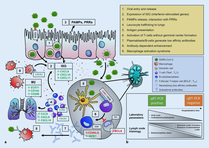
A dysregulated immune response is considered one of the major factors leading to severe COVID-19. Previously described mechanisms include the development of a cytokine storm, missing immunoglobulin class switch, antibody-mediated enhancement, and aberrant antigen presentation.

OBJECTIVES

To understand the heterogeneity of immune response in COVID-19, a thorough investigation of histomorphological patterns in regional lymph nodes was performed.

MATERIALS AND METHODS

Lymph nodes from the cervical, mediastinal, and hilar regions were extracted from autopsies of patients with lethal COVID-19 (n = 20). Histomorphological characteristics, SARS-CoV‑2 qRT-PCR, and gene expression profiling on common genes involved in immunologic response were analyzed.

RESULTS

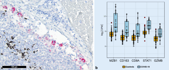
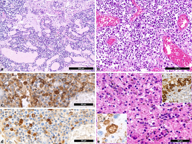
Lymph nodes displayed moderate to severe capillary stasis and edema, an increased presence of extrafollicular plasmablasts, mild to moderate plasmacytosis, a dominant population of CD8 T‑cells, and CD11c/CD68 histiocytosis with hemophagocytic activity. Out of 20 cases, 18 presented with hypoplastic or missing germinal centers with a decrease of follicular dendritic cells and follicular T‑helper cells. A positive viral load was detected by qRT-PCR in 14 of 20 cases, yet immunohistochemistry for SARS-CoV-2 N-antigen revealed positivity in sinus histiocytes of only one case. Gene expression analysis revealed an increased expression of STAT1, CD163, granzyme B, CD8A, MZB1, and PAK1, as well as CXCL9.

CONCLUSIONS

Taken together, our findings imply a dysregulated immune response in lethal COVID-19. The absence/hypoplasia of germinal centers and increased presence of plasmablasts implies a transient B‑cell response, implying an impaired development of long-term immunity against SARS-CoV‑2 in such occasions.

摘要

背景

免疫反应失调被认为是导致严重 COVID-19 的主要因素之一。先前描述的机制包括细胞因子风暴的发展、免疫球蛋白类别转换缺失、抗体介导的增强以及异常抗原呈递。

目的

为了了解 COVID-19 中免疫反应的异质性,对区域性淋巴结的组织形态模式进行了彻底调查。

材料和方法

从致死性 COVID-19 患者(n=20)的尸检中提取颈、纵隔和肺门区域的淋巴结。分析组织形态学特征、SARS-CoV-2 qRT-PCR 和常见免疫反应相关基因的基因表达谱。

结果

淋巴结显示中等至重度毛细血管淤滞和水肿,滤泡外浆母细胞增多,轻度至中度浆细胞增多,CD8 T 细胞占主导地位,CD11c/CD68 组织细胞伴有噬血细胞活性。在 20 例中,有 18 例表现为生发中心发育不良或缺失,滤泡树突状细胞和滤泡辅助性 T 细胞减少。qRT-PCR 检测到 14 例中有阳性病毒载量,但 SARS-CoV-2 N 抗原的免疫组化仅在 1 例中显示阳性。基因表达分析显示 STAT1、CD163、颗粒酶 B、CD8A、MZB1 和 PAK1 以及 CXCL9 的表达增加。

结论

综上所述,我们的发现表明致命性 COVID-19 中存在免疫反应失调。生发中心的缺失/发育不良和浆母细胞的增多暗示着短暂的 B 细胞反应,这意味着在这种情况下对 SARS-CoV-2 产生长期免疫的能力受损。

https://cdn.ncbi.nlm.nih.gov/pmc/blobs/cea7/8571157/4c4fb4dbfe48/292_2021_945_Fig3_HTML.jpg
https://cdn.ncbi.nlm.nih.gov/pmc/blobs/cea7/8571157/c93267c9e265/292_2021_945_Fig1_HTML.jpg
https://cdn.ncbi.nlm.nih.gov/pmc/blobs/cea7/8571157/2df76df761b9/292_2021_945_Fig2_HTML.jpg
https://cdn.ncbi.nlm.nih.gov/pmc/blobs/cea7/8571157/4c4fb4dbfe48/292_2021_945_Fig3_HTML.jpg
https://cdn.ncbi.nlm.nih.gov/pmc/blobs/cea7/8571157/c93267c9e265/292_2021_945_Fig1_HTML.jpg
https://cdn.ncbi.nlm.nih.gov/pmc/blobs/cea7/8571157/2df76df761b9/292_2021_945_Fig2_HTML.jpg
https://cdn.ncbi.nlm.nih.gov/pmc/blobs/cea7/8571157/4c4fb4dbfe48/292_2021_945_Fig3_HTML.jpg

相似文献

1
Histomorphological patterns of regional lymph nodes in COVID-19 lungs.COVID-19 肺部的区域性淋巴结的组织形态学模式。
Pathologe. 2021 Nov;42(Suppl 1):89-97. doi: 10.1007/s00292-021-00945-6. Epub 2021 May 5.
2
[Histomorphological patterns of regional lymph nodes in COVID-19 lungs].[新型冠状病毒肺炎肺部区域淋巴结的组织形态学模式]
Pathologe. 2021 Mar;42(2):188-196. doi: 10.1007/s00292-021-00914-z. Epub 2021 Feb 11.
3
Vascular Damage, Thromboinflammation, Plasmablast Activation, T-Cell Dysregulation and Pathological Histiocytic Response in Pulmonary Draining Lymph Nodes of COVID-19.COVID-19 肺部引流淋巴结中的血管损伤、血栓炎症、浆细胞激活、T 细胞失调和病理性组织细胞反应。
Front Immunol. 2021 Dec 13;12:763098. doi: 10.3389/fimmu.2021.763098. eCollection 2021.
4
Viral Load and Patterns of SARS-CoV-2 Dissemination to the Lungs, Mediastinal Lymph Nodes, and Spleen of Patients with COVID-19 Associated Lymphopenia.COVID-19 相关淋巴细胞减少症患者的病毒载量及 SARS-CoV-2 向肺部、纵隔淋巴结和脾脏传播的模式。
Viruses. 2021 Jul 20;13(7):1410. doi: 10.3390/v13071410.
5
Deficiency of Tfh Cells and Germinal Center in Deceased COVID-19 Patients.新冠肺炎死者体内 Tfh 细胞和生发中心缺失。
Curr Med Sci. 2020 Aug;40(4):618-624. doi: 10.1007/s11596-020-2225-x. Epub 2020 Aug 29.
6
Loss of Bcl-6-Expressing T Follicular Helper Cells and Germinal Centers in COVID-19.新冠病毒(COVID-19)中 Bcl-6 表达的滤泡辅助性 T 细胞和生发中心的缺失。
Cell. 2020 Oct 1;183(1):143-157.e13. doi: 10.1016/j.cell.2020.08.025. Epub 2020 Aug 19.
7
Immune Response to SARS-CoV-2 Vaccine in 2 Men.两例男性对 SARS-CoV-2 疫苗的免疫反应。
Int Arch Allergy Immunol. 2022;183(3):350-359. doi: 10.1159/000520046. Epub 2021 Nov 18.
8
Low Innate Immunity and Lagged Adaptive Immune Response in the Re-Tested Viral RNA Positivity of a COVID-19 Patient.COVID-19 患者再次检测到病毒 RNA 阳性时存在低固有免疫和滞后适应性免疫反应。
Front Immunol. 2021 Jul 1;12:664619. doi: 10.3389/fimmu.2021.664619. eCollection 2021.
9
Comparison of RNA In Situ Hybridization and Immunohistochemistry Techniques for the Detection and Localization of SARS-CoV-2 in Human Tissues.RNA 原位杂交与免疫组织化学技术在检测和定位人组织中 SARS-CoV-2 的比较。
Am J Surg Pathol. 2021 Jan;45(1):14-24. doi: 10.1097/PAS.0000000000001563.
10
Novel insight from the first lung transplant of a COVID-19 patient.首例 COVID-19 患者肺移植的新见解。
Eur J Clin Invest. 2021 Jan;51(1):e13443. doi: 10.1111/eci.13443. Epub 2020 Nov 20.

引用本文的文献

1
Potential Immune-Inflammatory Proteome Biomarkers for Guiding the Treatment of Patients with Primary Acute Angle-Closure Glaucoma Caused by COVID-19.用于指导 COVID-19 引起的原发性急性闭角型青光眼患者治疗的潜在免疫炎症蛋白质组生物标志物。
J Proteome Res. 2024 Jul 5;23(7):2587-2597. doi: 10.1021/acs.jproteome.4c00325. Epub 2024 Jun 5.
2
Sarcoidosis-related autoimmune inflammation in COVID-19 convalescent patients.新冠康复患者中与结节病相关的自身免疫性炎症
Front Med (Lausanne). 2023 Dec 21;10:1271198. doi: 10.3389/fmed.2023.1271198. eCollection 2023.
3
B-cell and antibody responses to SARS-CoV-2: infection, vaccination, and hybrid immunity.

本文引用的文献

1
Haemophagocytic lymphohistiocytosis and liver failure-induced massive hyperferritinaemia in a male COVID-19 patient.一名男性 COVID-19 患者发生噬血细胞性淋巴组织细胞增生症和肝衰竭引起的大量血铁蛋白血症。
Swiss Med Wkly. 2021 Jan 15;151:w20420. doi: 10.4414/smw.2021.20420. eCollection 2021 Jan 18.
2
Switched and unswitched memory B cells detected during SARS-CoV-2 convalescence correlate with limited symptom duration.在 SARS-CoV-2 恢复期检测到的已转换和未转换记忆 B 细胞与症状持续时间有限相关。
PLoS One. 2021 Jan 28;16(1):e0244855. doi: 10.1371/journal.pone.0244855. eCollection 2021.
3
Individuals With Less Severe Manifestations of SARS-CoV-2 Infection May Not Develop Long-Lasting Humoral Immunity.
B 细胞和抗体对 SARS-CoV-2 的反应:感染、疫苗接种和混合免疫。
Cell Mol Immunol. 2024 Feb;21(2):144-158. doi: 10.1038/s41423-023-01095-w. Epub 2023 Nov 10.
4
The Janus face of proliferating plasmablasts in dengue and COVID-19 infections.登革热和 COVID-19 感染中增殖性浆母细胞的双面性。
Front Immunol. 2023 Aug 11;14:1068424. doi: 10.3389/fimmu.2023.1068424. eCollection 2023.
5
Unexpected CD5 B Cell Lymphocytosis during SARS-CoV-2 Infection: Relevance for the Pathophysiology of Chronic Lymphocytic Leukemia.新型冠状病毒感染期间意外出现的CD5 B细胞淋巴细胞增多症:与慢性淋巴细胞白血病病理生理学的相关性
J Clin Med. 2023 Jan 28;12(3):998. doi: 10.3390/jcm12030998.
6
Reconstructed Genome-Scale Metabolic Model Characterizes Adaptive Metabolic Flux Changes in Peripheral Blood Mononuclear Cells in Severe COVID-19 Patients.重建的基因组规模代谢模型描绘了严重 COVID-19 患者外周血单核细胞中的适应性代谢通量变化。
Int J Mol Sci. 2022 Oct 17;23(20):12400. doi: 10.3390/ijms232012400.
7
SARS-CoV-2 Brain Regional Detection, Histopathology, Gene Expression, and Immunomodulatory Changes in Decedents with COVID-19.COVID-19 逝者的 SARS-CoV-2 脑部区域检测、组织病理学、基因表达和免疫调节变化。
J Neuropathol Exp Neurol. 2022 Aug 16;81(9):666-695. doi: 10.1093/jnen/nlac056.
8
Tissue immunity to SARS-CoV-2: Role in protection and immunopathology.组织对 SARS-CoV-2 的免疫:在保护和免疫病理中的作用。
Immunol Rev. 2022 Aug;309(1):25-39. doi: 10.1111/imr.13112. Epub 2022 Jun 25.
9
Immune imprinting, breadth of variant recognition, and germinal center response in human SARS-CoV-2 infection and vaccination.人类 SARS-CoV-2 感染和接种疫苗中的免疫印迹、变体识别广度和生发中心反应。
Cell. 2022 Mar 17;185(6):1025-1040.e14. doi: 10.1016/j.cell.2022.01.018. Epub 2022 Jan 25.
10
COVID-19: Multiorgan Dissemination of SARS-CoV-2 Is Driven by Pulmonary Factors.COVID-19:SARS-CoV-2 的多器官传播由肺部因素驱动。
Viruses. 2021 Dec 26;14(1):39. doi: 10.3390/v14010039.
感染新冠病毒(SARS-CoV-2)症状较轻的个体可能不会产生持久的体液免疫。
Am J Clin Pathol. 2021 Feb 4;155(2):320. doi: 10.1093/ajcp/aqaa233.
4
Two distinct immunopathological profiles in autopsy lungs of COVID-19.COVID-19 患者尸检肺组织中的两种不同免疫病理特征。
Nat Commun. 2020 Oct 8;11(1):5086. doi: 10.1038/s41467-020-18854-2.
5
Toll-like receptors and COVID-19: a two-faced story with an exciting ending.Toll样受体与新型冠状病毒肺炎:一个有着激动人心结局的双面故事。
Future Sci OA. 2020 Jul 30;6(8):FSO605. doi: 10.2144/fsoa-2020-0091.
6
Autoantibodies against type I IFNs in patients with life-threatening COVID-19.COVID-19 危重症患者体内针对 I 型干扰素的自身抗体。
Science. 2020 Oct 23;370(6515). doi: 10.1126/science.abd4585. Epub 2020 Sep 24.
7
Inborn errors of type I IFN immunity in patients with life-threatening COVID-19.COVID-19 危重症患者的 I 型 IFN 免疫先天缺陷。
Science. 2020 Oct 23;370(6515). doi: 10.1126/science.abd4570. Epub 2020 Sep 24.
8
Biomarkers and outcomes of COVID-19 hospitalisations: systematic review and meta-analysis.COVID-19 住院患者的生物标志物和结局:系统评价和荟萃分析。
BMJ Evid Based Med. 2021 Jun;26(3):107-108. doi: 10.1136/bmjebm-2020-111536. Epub 2020 Sep 15.
9
Loss of Bcl-6-Expressing T Follicular Helper Cells and Germinal Centers in COVID-19.新冠病毒(COVID-19)中 Bcl-6 表达的滤泡辅助性 T 细胞和生发中心的缺失。
Cell. 2020 Oct 1;183(1):143-157.e13. doi: 10.1016/j.cell.2020.08.025. Epub 2020 Aug 19.
10
Adaptive immune responses to SARS-CoV-2 infection in severe versus mild individuals.严重与轻度个体对 SARS-CoV-2 感染的适应性免疫反应。
Signal Transduct Target Ther. 2020 Aug 14;5(1):156. doi: 10.1038/s41392-020-00263-y.