• 文献检索
  • 文档翻译
  • 深度研究
  • 学术资讯
  • Suppr Zotero 插件Zotero 插件
  • 邀请有礼
  • 套餐&价格
  • 历史记录
应用&插件
Suppr Zotero 插件Zotero 插件浏览器插件Mac 客户端Windows 客户端微信小程序
定价
高级版会员购买积分包购买API积分包
服务
文献检索文档翻译深度研究API 文档MCP 服务
关于我们
关于 Suppr公司介绍联系我们用户协议隐私条款
关注我们

Suppr 超能文献

核心技术专利:CN118964589B侵权必究
粤ICP备2023148730 号-1Suppr @ 2026

文献检索

告别复杂PubMed语法,用中文像聊天一样搜索,搜遍4000万医学文献。AI智能推荐,让科研检索更轻松。

立即免费搜索

文件翻译

保留排版,准确专业,支持PDF/Word/PPT等文件格式,支持 12+语言互译。

免费翻译文档

深度研究

AI帮你快速写综述,25分钟生成高质量综述,智能提取关键信息,辅助科研写作。

立即免费体验

脊髓小脑性共济失调 3 型的区域性脑和脊髓体积损失。

Regional Brain and Spinal Cord Volume Loss in Spinocerebellar Ataxia Type 3.

机构信息

DZNE, German Center for Neurodegenerative Diseases, Bonn, Germany.

Department of Neurology, University Hospital Bonn, Bonn, Germany.

出版信息

Mov Disord. 2021 Oct;36(10):2273-2281. doi: 10.1002/mds.28610. Epub 2021 May 5.

DOI:10.1002/mds.28610
PMID:33951232
原文链接:https://pmc.ncbi.nlm.nih.gov/articles/PMC9521507/
Abstract

BACKGROUND

Given that new therapeutic options for spinocerebellar ataxias are on the horizon, there is a need for markers that reflect disease-related alterations, in particular, in the preataxic stage, in which clinical scales are lacking sensitivity.

OBJECTIVE

The objective of this study was to quantify regional brain volumes and upper cervical spinal cord areas in spinocerebellar ataxia type 3 in vivo across the entire time course of the disease.

METHODS

We applied a brain segmentation approach that included a lobular subsegmentation of the cerebellum to magnetic resonance images of 210 ataxic and 48 preataxic spinocerebellar ataxia type 3 mutation carriers and 63 healthy controls. In addition, cervical cord cross-sectional areas were determined at 2 levels.

RESULTS

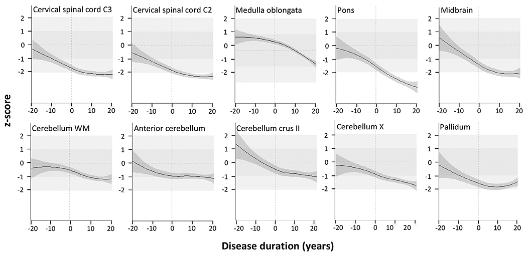
The metrics of cervical spinal cord segments C3 and C2, medulla oblongata, pons, and pallidum, and the cerebellar anterior lobe were reduced in preataxic mutation carriers compared with controls. Those of cervical spinal cord segments C2 and C3, medulla oblongata, pons, midbrain, cerebellar lobules crus II and X, cerebellar white matter, and pallidum were reduced in ataxic compared with nonataxic carriers. Of all metrics studied, pontine volume showed the steepest decline across the disease course. It covaried with ataxia severity, CAG repeat length, and age. The multivariate model derived from this analysis explained 46.33% of the variance of pontine volume.

CONCLUSION

Regional brain and spinal cord tissue loss in spinocerebellar ataxia type 3 starts before ataxia onset. Pontine volume appears to be the most promising imaging biomarker candidate for interventional trials that aim at slowing the progression of spinocerebellar ataxia type 3. © 2021 The Authors. Movement Disorders published by Wiley Periodicals LLC on behalf of International Parkinson and Movement Disorder Society.

摘要

背景

鉴于新的脊髓小脑共济失调治疗选择即将出现,因此需要有反映疾病相关变化的标志物,特别是在临床量表缺乏敏感性的前期阶段。

目的

本研究旨在定量分析脊髓小脑共济失调 3 型患者在整个疾病过程中的脑区体积和上颈段脊髓面积。

方法

我们应用了一种脑部分割方法,该方法包括小脑的小叶亚分割,对 210 例共济失调和 48 例前期脊髓小脑共济失调 3 型突变携带者以及 63 名健康对照者的磁共振成像进行了分析。此外,还在 2 个水平上确定了颈段脊髓的横截面积。

结果

与对照组相比,前期突变携带者的颈段脊髓 C3 和 C2 节段、延髓、脑桥和苍白球以及小脑前叶的测量值均减小。与非共济失调携带者相比,颈段脊髓 C2 和 C3 节段、延髓、脑桥、中脑、小脑小叶 crus II 和 X、小脑白质和苍白球的测量值均减小。在所研究的所有指标中,脑桥体积在整个疾病过程中的下降幅度最大。它与共济失调严重程度、CAG 重复长度和年龄相关。该分析得出的多元模型解释了脑桥体积变化的 46.33%。

结论

脊髓小脑共济失调 3 型患者的脑和脊髓组织损失在发病前就开始了。脑桥体积似乎是最有前途的影像学生物标志物候选者,可用于旨在减缓脊髓小脑共济失调 3 型进展的干预性试验。© 2021 作者。运动障碍由 Wiley 期刊公司代表国际帕金森病和运动障碍协会出版。

https://cdn.ncbi.nlm.nih.gov/pmc/blobs/00ce/9521507/a1d8b56f49c0/nihms-1837147-f0001.jpg

相似文献

1
Regional Brain and Spinal Cord Volume Loss in Spinocerebellar Ataxia Type 3.脊髓小脑性共济失调 3 型的区域性脑和脊髓体积损失。
Mov Disord. 2021 Oct;36(10):2273-2281. doi: 10.1002/mds.28610. Epub 2021 May 5.
2
The Pattern and Stages of Atrophy in Spinocerebellar Ataxia Type 2: Volumetrics from ENIGMA-Ataxia.2型脊髓小脑共济失调萎缩的模式与阶段:来自ENIGMA-共济失调研究的容积分析
Mov Disord. 2025 Apr;40(4):651-661. doi: 10.1002/mds.30143. Epub 2025 Feb 10.
3
A 5-Year Longitudinal Clinical and Magnetic Resonance Imaging Study in Spinocerebellar Ataxia Type 3.脊髓小脑性共济失调 3 型的 5 年纵向临床和磁共振成像研究。
Mov Disord. 2020 Sep;35(9):1679-1684. doi: 10.1002/mds.28113. Epub 2020 Jun 9.
4
Longitudinal Changes of Clinical, Imaging, and Fluid Biomarkers in Preataxic and Early Ataxic Spinocerebellar Ataxia Type 2 and 7 Carriers.未病期和早期小脑共济失调型 2 型和 7 型脊髓小脑共济失调携带者的临床、影像学和液体生物标志物的纵向变化。
Neurology. 2024 Sep 10;103(5):e209749. doi: 10.1212/WNL.0000000000209749. Epub 2024 Aug 12.
5
Progression of Clinical and Eye Movement Markers in Preataxic Carriers of Machado-Joseph Disease.马查多-约瑟夫病共济失调前期携带者的临床及眼动标志物进展
Mov Disord. 2023 Jan;38(1):26-34. doi: 10.1002/mds.29226. Epub 2022 Sep 21.
6
Genotype-specific spinal cord damage in spinocerebellar ataxias: an ENIGMA-Ataxia study.脊髓小脑共济失调中基因型特异性脊髓损伤:一项 ENIGMA-Ataxia 研究。
J Neurol Neurosurg Psychiatry. 2024 Jun 17;95(7):682-690. doi: 10.1136/jnnp-2023-332696.
7
Genotype-specific patterns of atrophy progression are more sensitive than clinical decline in SCA1, SCA3 and SCA6.特定基因型的萎缩进展模式比 SCA1、SCA3 和 SCA6 的临床衰退更敏感。
Brain. 2013 Mar;136(Pt 3):905-17. doi: 10.1093/brain/aws369. Epub 2013 Feb 18.
8
Baseline Clinical and Blood Biomarkers in Patients With Preataxic and Early-Stage Disease Spinocerebellar Ataxia 1 and 3.脊髓小脑性共济失调 1 型和 3 型早前期和早期疾病患者的基线临床和血液生物标志物。
Neurology. 2023 Apr 25;100(17):e1836-e1848. doi: 10.1212/WNL.0000000000207088. Epub 2023 Feb 16.
9
Cervical Spinal Cord Degeneration in Spinocerebellar Ataxia Type 7.脊髓小脑共济失调7型中的颈脊髓退变
AJNR Am J Neuroradiol. 2021 Sep;42(9):1735-1739. doi: 10.3174/ajnr.A7202. Epub 2021 Jul 1.
10
Spinal Cord Damage in Spinocerebellar Ataxia Type 1.脊髓小脑共济失调1型中的脊髓损伤
Cerebellum. 2017 Aug;16(4):792-796. doi: 10.1007/s12311-017-0854-9.

引用本文的文献

1
Altered Brain Iron Depositions of Spinocerebellar Ataxia Type 3: From Pre-Symptomatic to Symptomatic Stage.3型脊髓小脑共济失调患者脑铁沉积的变化:从症状前阶段到症状阶段
Eur J Neurol. 2025 Apr;32(4):e70127. doi: 10.1111/ene.70127.
2
Associations between CAG repeat size, brain and spinal cord volume loss, and motor symptoms in spinocerebellar ataxia type 3: a cohort study.3型脊髓小脑共济失调中CAG重复序列长度、脑和脊髓体积丢失与运动症状之间的关联:一项队列研究
Orphanet J Rare Dis. 2025 Jan 23;20(1):35. doi: 10.1186/s13023-025-03531-8.
3
Longitudinal Changes of Clinical, Imaging, and Fluid Biomarkers in Preataxic and Early Ataxic Spinocerebellar Ataxia Type 2 and 7 Carriers.

本文引用的文献

1
Conversion of individuals at risk for spinocerebellar ataxia types 1, 2, 3, and 6 to manifest ataxia (RISCA): a longitudinal cohort study.将脊髓小脑共济失调 1、2、3、6 型的风险个体转化为显性共济失调(RISCA):一项纵向队列研究。
Lancet Neurol. 2020 Sep;19(9):738-747. doi: 10.1016/S1474-4422(20)30235-0.
2
Longitudinal analysis of regional cerebellum volumes during normal aging.正常衰老过程中小脑区域体积的纵向分析。
Neuroimage. 2020 Oct 15;220:117062. doi: 10.1016/j.neuroimage.2020.117062. Epub 2020 Jun 25.
3
A 5-Year Longitudinal Clinical and Magnetic Resonance Imaging Study in Spinocerebellar Ataxia Type 3.
未病期和早期小脑共济失调型 2 型和 7 型脊髓小脑共济失调携带者的临床、影像学和液体生物标志物的纵向变化。
Neurology. 2024 Sep 10;103(5):e209749. doi: 10.1212/WNL.0000000000209749. Epub 2024 Aug 12.
4
Sensitivity of Advanced Magnetic Resonance Imaging to Progression over Six Months in Early Spinocerebellar Ataxia.高级磁共振成像对早期脊髓小脑共济失调 6 个月进展的敏感性。
Mov Disord. 2024 Oct;39(10):1856-1867. doi: 10.1002/mds.29934. Epub 2024 Jul 26.
5
Genotype-specific spinal cord damage in spinocerebellar ataxias: an ENIGMA-Ataxia study.脊髓小脑共济失调中基因型特异性脊髓损伤:一项 ENIGMA-Ataxia 研究。
J Neurol Neurosurg Psychiatry. 2024 Jun 17;95(7):682-690. doi: 10.1136/jnnp-2023-332696.
6
Cerebellar Volumetry in Ataxias: Relation to Ataxia Severity and Duration.小脑容积测量在共济失调中的应用:与共济失调严重程度和持续时间的关系。
Cerebellum. 2024 Aug;23(4):1521-1529. doi: 10.1007/s12311-024-01659-0. Epub 2024 Feb 16.
7
On the Cut-Off Value of the Anteroposterior Diameter of the Midbrain Atrophy in Spinocerebellar Ataxia Type 2 Patients.2型脊髓小脑共济失调患者中脑萎缩前后径的截断值研究
Brain Sci. 2024 Jan 5;14(1):53. doi: 10.3390/brainsci14010053.
8
Early Neuroaxonal Damage in Neurologic Disorders Associated With GAD65 Antibodies.神经节苷脂抗体相关神经紊乱的早期神经轴突损伤。
Neurol Neuroimmunol Neuroinflamm. 2023 Nov 1;11(1). doi: 10.1212/NXI.0000000000200176. Print 2024 Jan.
9
Disease progression of spinocerebellar ataxia types 1, 2, 3 and 6 before and after ataxia onset.脊髓小脑共济失调 1、2、3 和 6 型在共济失调发作前后的疾病进展。
Ann Clin Transl Neurol. 2023 Oct;10(10):1833-1843. doi: 10.1002/acn3.51875. Epub 2023 Aug 17.
10
The confound of hemodynamic response function variability in human resting-state functional MRI studies.人类静息态功能磁共振成像研究中血液动力学响应函数变异性的混杂因素。
Front Neurosci. 2023 Jul 14;17:934138. doi: 10.3389/fnins.2023.934138. eCollection 2023.
脊髓小脑性共济失调 3 型的 5 年纵向临床和磁共振成像研究。
Mov Disord. 2020 Sep;35(9):1679-1684. doi: 10.1002/mds.28113. Epub 2020 Jun 9.
4
Prediction of Survival With Long-Term Disease Progression in Most Common Spinocerebellar Ataxia.最常见的脊髓小脑共济失调的长期疾病进展的生存预测。
Mov Disord. 2019 Aug;34(8):1220-1227. doi: 10.1002/mds.27739. Epub 2019 Jun 18.
5
Unique degeneration signatures in the cerebellar cortex for spinocerebellar ataxias 2, 3, and 7.脊髓小脑共济失调 2、3 和 7 型小脑皮质的独特退化特征。
Neuroimage Clin. 2018;20:931-938. doi: 10.1016/j.nicl.2018.09.026. Epub 2018 Sep 27.
6
Structural signature of SCA3: From presymptomatic to late disease stages.SCA3 的结构特征:从无症状期到疾病晚期。
Ann Neurol. 2018 Sep;84(3):401-408. doi: 10.1002/ana.25297. Epub 2018 Sep 4.
7
Autosomal dominant cerebellar ataxias: Imaging biomarkers with high effect sizes.常染色体显性小脑共济失调:具有高效应量的影像学生物标志物。
Neuroimage Clin. 2018 Jun 14;19:858-867. doi: 10.1016/j.nicl.2018.06.011. eCollection 2018.
8
Oligonucleotide therapy mitigates disease in spinocerebellar ataxia type 3 mice.寡核苷酸疗法减轻脊髓小脑共济失调 3 型小鼠的疾病。
Ann Neurol. 2018 Jul;84(1):64-77. doi: 10.1002/ana.25264. Epub 2018 Aug 6.
9
Brain atrophy measures in preclinical and manifest spinocerebellar ataxia type 2.临床前和明显的2型脊髓小脑共济失调的脑萎缩测量
Ann Clin Transl Neurol. 2018 Jan 7;5(2):128-137. doi: 10.1002/acn3.504. eCollection 2018 Feb.
10
The Neuropathology of Spinocerebellar Ataxia Type 3/Machado-Joseph Disease.脊髓小脑共济失调 3 型/马查多-约瑟夫病的神经病理学。
Adv Exp Med Biol. 2018;1049:233-241. doi: 10.1007/978-3-319-71779-1_11.