Cardiovascular Branch, Division of Intramural Research, National Heart Lung and Blood Institute, National Institutes of Health, Bethesda, MD, 20892, USA.
Division of Cardiology, Department of Medicine, Johns Hopkins University School of Medicine, Baltimore, MD, USA.
J Cardiovasc Magn Reson. 2021 May 6;23(1):50. doi: 10.1186/s12968-020-00693-1.
Low-field (0.55 T) high-performance cardiovascular magnetic resonance (CMR) is an attractive platform for CMR-guided intervention as device heating is reduced around 7.5-fold compared to 1.5 T. This work determines the feasibility of visualizing cardiac radiofrequency (RF) ablation lesions at low field CMR and explores a novel alternative method for targeted tissue destruction: acetic acid chemoablation.
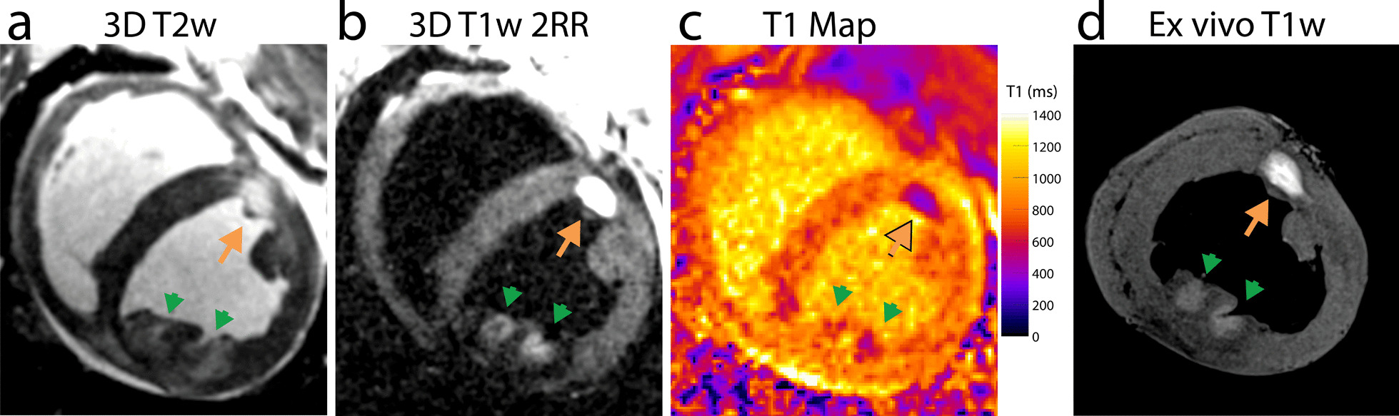
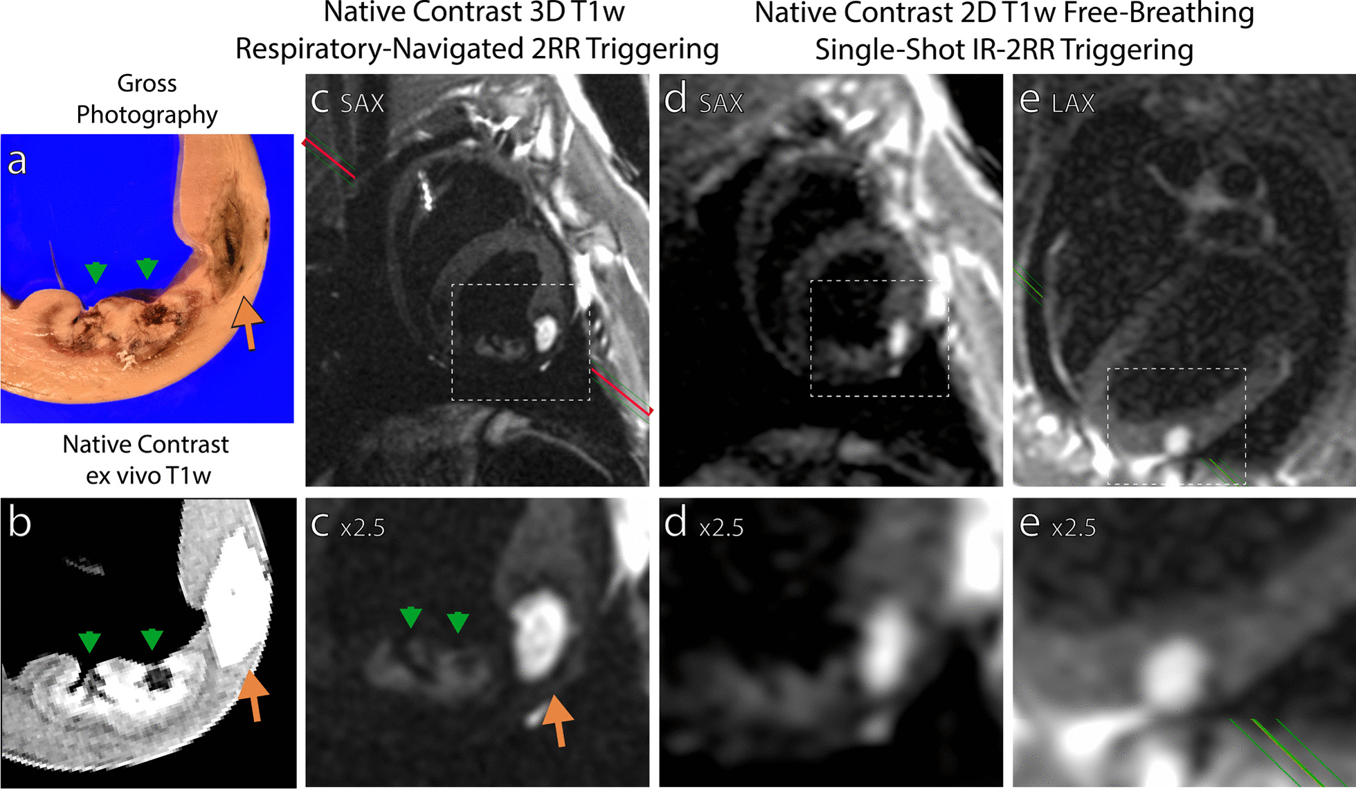
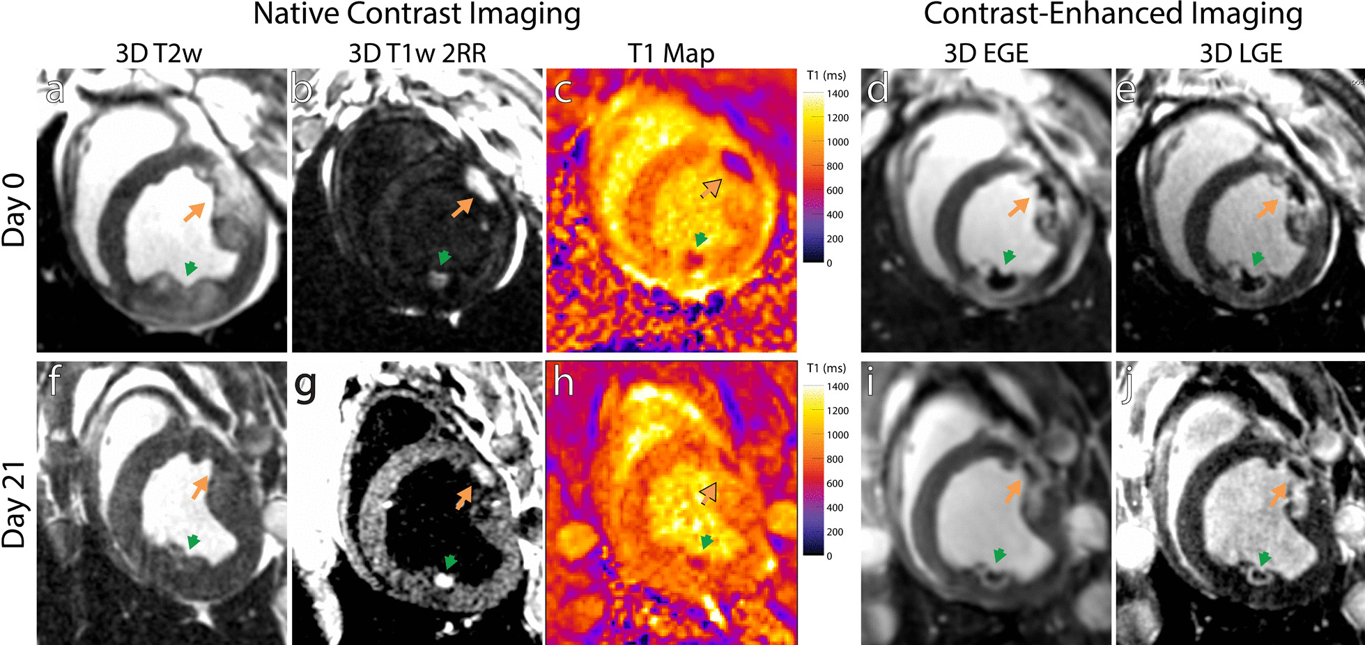
N = 10 swine underwent X-ray fluoroscopy-guided RF ablation (6-7 lesions) and acetic acid chemoablation (2-3 lesions) of the left ventricle. Animals were imaged at 0.55 T with native contrast 3D-navigator gated T1-weighted T1w) CMR for lesion visualization, gated single-shot imaging to determine potential for real-time visualization of lesion formation, and T1 mapping to measure change in T1 in response to ablation. Seven animals were euthanized on ablation day and hearts imaged ex vivo. The remaining animals were imaged again in vivo at 21 days post ablation to observe lesion evolution.
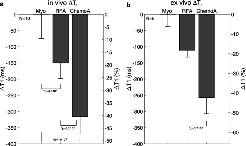
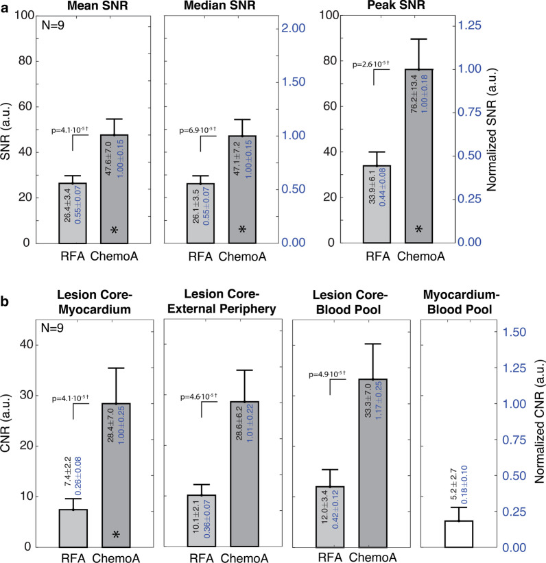
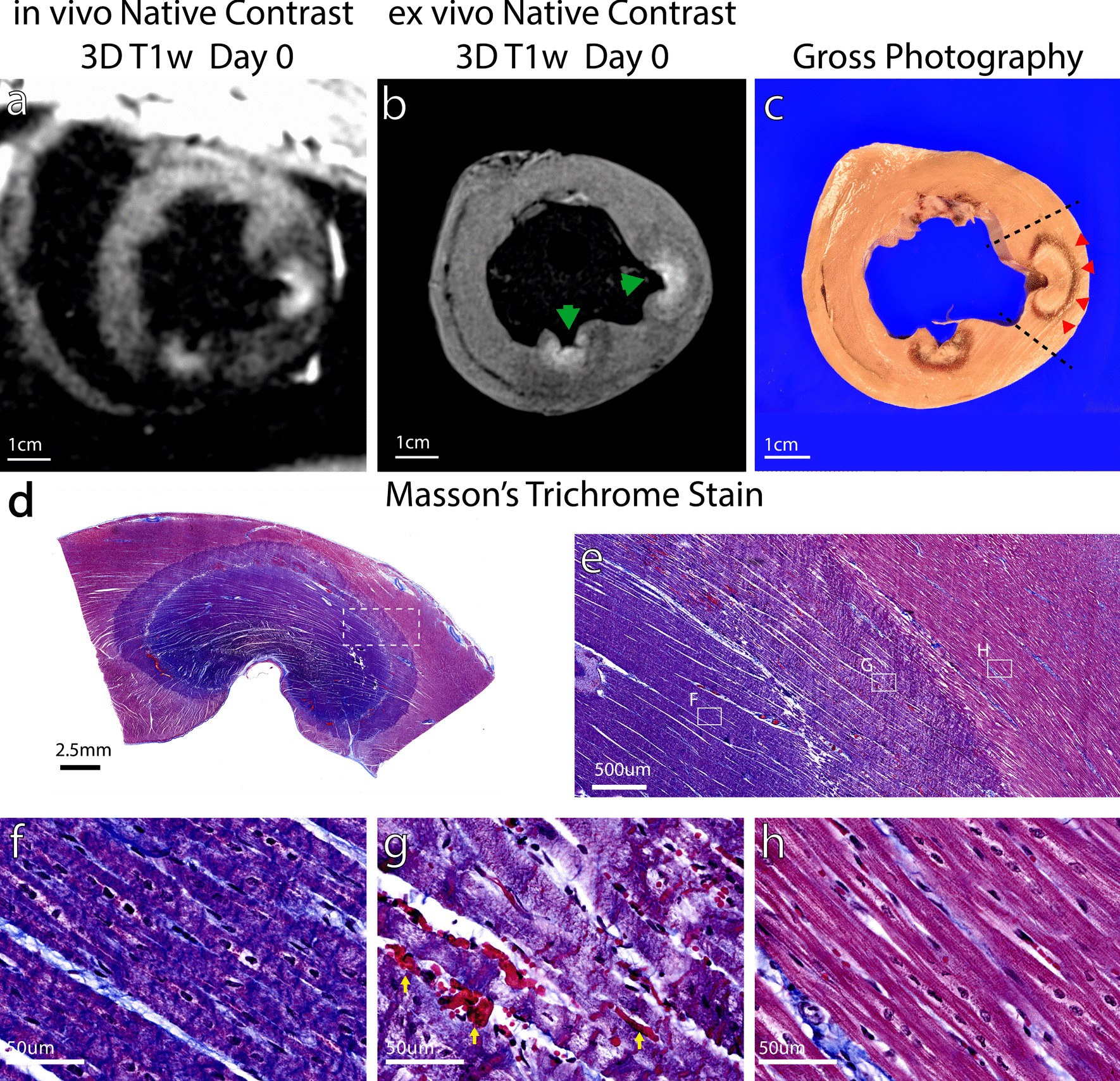
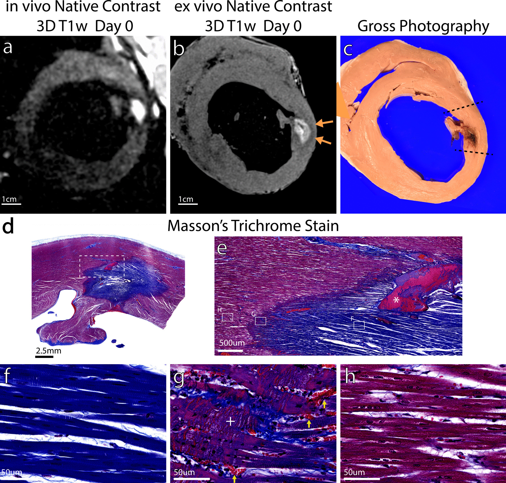
Chemoablation lesions could be visualized and displayed much higher contrast than necrotic RF ablation lesions with T1w imaging. On the day of ablation, in vivo myocardial T1 dropped by 19 ± 7% in RF ablation lesion cores, and by 40 ± 7% in chemoablation lesion cores (p < 4e-5). In high resolution ex vivo imaging, with reduced partial volume effects, lesion core T1 dropped by 18 ± 3% and 42 ± 6% for RF and chemoablation, respectively. Mean, median, and peak lesion signal-to-noise ratio (SNR) were all at least 75% higher with chemoablation. Lesion core to myocardium contrast-to-noise (CNR) was 3.8 × higher for chemoablation. Correlation between in vivo and ex vivo CMR and histology indicated that the periphery of RF ablation lesions do not exhibit changes in T1 while the entire extent of chemoablation exhibits T1 changes. Correlation of T1w enhancing lesion volumes indicated in vivo estimates of lesion volume are accurate for chemoablation but underestimate extent of necrosis for RF ablation.
The visualization of coagulation necrosis from cardiac ablation is feasible using low-field high-performance CMR. Chemoablation produced a more pronounced change in lesion T1 than RF ablation, increasing SNR and CNR and thereby making it easier to visualize in both 3D navigator-gated and real-time CMR and more suitable for low-field imaging.
与 1.5T 相比,低场(0.55T)高性能心脏磁共振(CMR)可显著减少设备加热,因此是 CMR 引导介入治疗的理想平台。本研究旨在确定在低场 CMR 下可视化心脏射频(RF)消融损伤的可行性,并探索一种新的靶向组织破坏方法:醋酸化学消融。
10 头猪接受 X 射线透视引导下 RF 消融(左心室 6-7 个病灶)和醋酸化学消融(左心室 2-3 个病灶)。动物在 0.55T 下采用原生对比度 3D 导航门控 T1 加权 T1w)CMR 进行成像,门控单次激发成像以确定实时可视化病变形成的可能性,以及 T1 映射以测量消融后 T1 的变化。7 只动物在消融当天处死,并进行心脏离体成像。其余动物在消融后 21 天再次进行体内成像,以观察病变演变。
T1w 成像可显示化学消融损伤,与坏死的 RF 消融损伤相比,对比度更高。在消融当天,RF 消融损伤核心的心肌 T1 降低了 19±7%,化学消融损伤核心的心肌 T1 降低了 40±7%(p<4e-5)。在高分辨率离体成像中,由于部分容积效应降低,RF 和化学消融的损伤核心 T1 分别降低了 18±3%和 42±6%。化学消融的平均、中位数和峰值病变信噪比(SNR)均至少提高了 75%。化学消融的病变核心与心肌的对比噪声比(CNR)高 3.8 倍。体内和离体 CMR 与组织学之间的相关性表明,RF 消融损伤的周边区域 T1 无变化,而整个化学消融区域 T1 均有变化。T1w 增强病变体积的相关性表明,化学消融的体内病变体积估计值准确,但 RF 消融的坏死程度估计值低估。
使用低场高性能 CMR 可以实现心脏消融后凝固性坏死的可视化。与 RF 消融相比,化学消融导致病变 T1 变化更明显,从而提高了 SNR 和 CNR,使病变在 3D 导航门控和实时 CMR 中更容易可视化,并更适合低场成像。