Ghafoori Elyar, Kholmovski Eugene G, Thomas Sam, Silvernagel Josh, Angel Nathan, Hu Nan, Dosdall Derek J, MacLeod Rob, Ranjan Ravi
From the Department of Bioengineering (E.G., J.S., N.A., D.J.D., R.M., R.R.), Cardiovascular Medicine (E.G., J.S., N.A., R.R.), UCAIR, Department of Radiology and Imaging Sciences (E.G.K.), Department of Medicine (S.T., N.H., R.R.), and Department of Surgery (D.J.D.), University of Utah, Salt Lake City.
Circ Arrhythm Electrophysiol. 2017 Nov;10(11). doi: 10.1161/CIRCEP.117.005599.
Magnetic resonance imaging (MRI) has been used to acutely visualize radiofrequency ablation lesions, but its accuracy in predicting chronic lesion size is unknown. The main goal of this study was to characterize different areas of enhancement in late gadolinium enhancement MRI done immediately after ablation to predict acute edema and chronic lesion size.
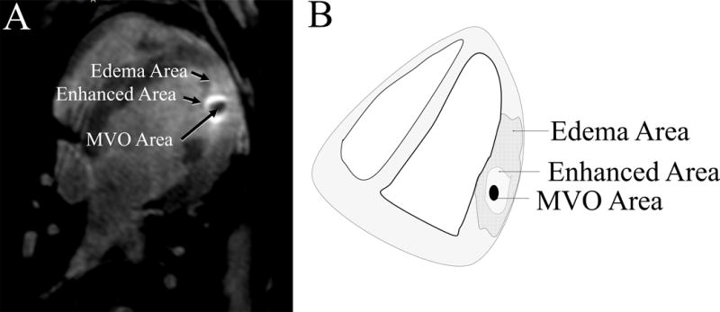
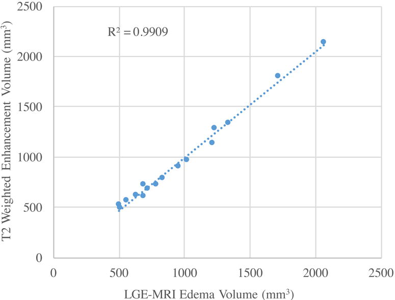
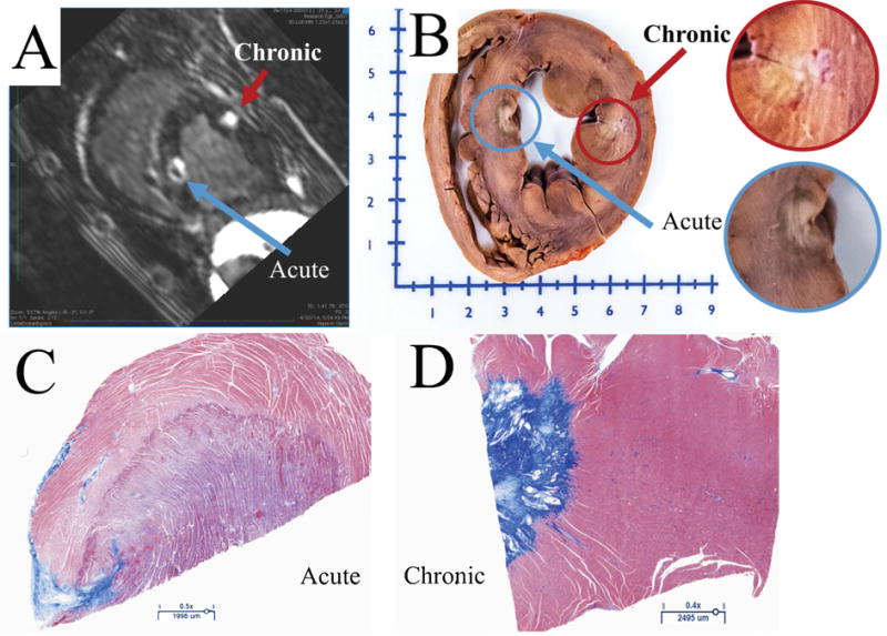
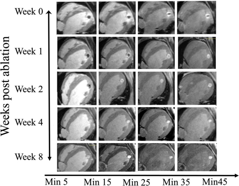
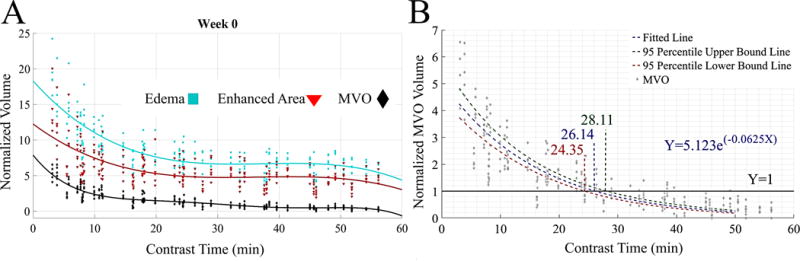
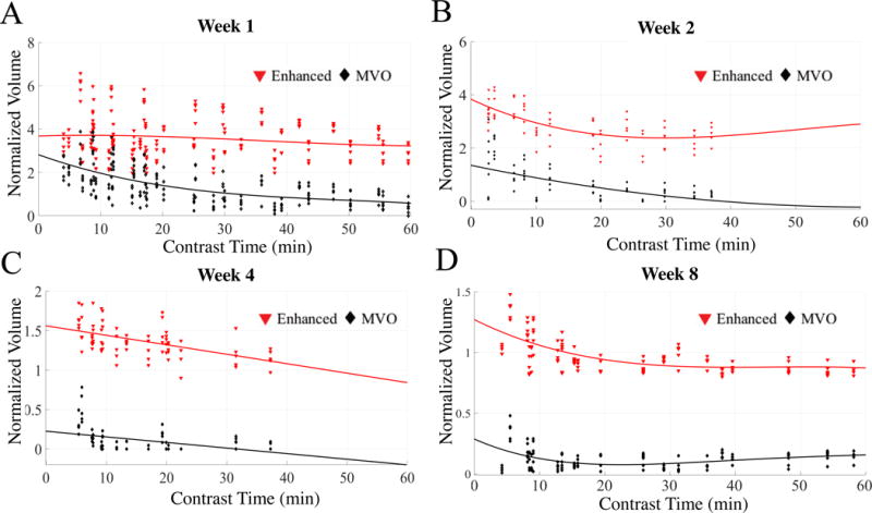
In a canine model (n=10), ventricular radiofrequency lesions were created using ThermoCool SmartTouch (Biosense Webster) catheter. All animals underwent MRI (late gadolinium enhancement and T2-weighted edema imaging) immediately after ablation and after 1, 2, 4, and 8 weeks. Edema, microvascular obstruction, and enhanced volumes were identified in MRI and normalized to chronic histological volume. Immediately after contrast administration, the microvascular obstruction region was 3.2±1.1 times larger than the chronic lesion volume in acute MRI. Even 60 minutes after contrast administration, edema was 8.7±3.31 times and the enhanced area 6.14±2.74 times the chronic lesion volume. Exponential fit to the microvascular obstruction volume was found to be the best predictor of chronic lesion volume at 26.14 minutes (95% prediction interval, 24.35-28.11 minutes) after contrast injection. The edema volume in late gadolinium enhancement correlated well with edema volume in T2-weighted MRI with an of 0.99.
Microvascular obstruction region on acute late gadolinium enhancement images acquired 26.1 minutes after contrast administration can accurately predict the chronic lesion volume. We also show that T1-weighted MRI images acquired immediately after contrast injection accurately shows edema resulting from radiofrequency ablation.
磁共振成像(MRI)已被用于急性可视化射频消融病灶,但其预测慢性病灶大小的准确性尚不清楚。本研究的主要目的是在消融后立即进行的延迟钆增强MRI中,对不同的强化区域进行特征分析,以预测急性水肿和慢性病灶大小。
在犬模型(n = 10)中,使用ThermoCool SmartTouch(Biosense Webster)导管制造心室射频病灶。所有动物在消融后立即以及1、2、4和8周后均接受MRI检查(延迟钆增强和T2加权水肿成像)。在MRI中识别出水肿、微血管阻塞和强化体积,并将其与慢性组织学体积进行归一化。在注射造影剂后立即,急性MRI中的微血管阻塞区域比慢性病灶体积大3.2±1.1倍。即使在注射造影剂60分钟后,水肿仍为慢性病灶体积的8.7±3.31倍,强化区域为慢性病灶体积的6.14±2.74倍。发现对微血管阻塞体积进行指数拟合是造影剂注射后26.14分钟(95%预测区间,24.35 - 28.11分钟)时慢性病灶体积的最佳预测指标。延迟钆增强中的水肿体积与T2加权MRI中的水肿体积相关性良好,相关系数为0.99。
在注射造影剂后第26.1分钟获得的急性延迟钆增强图像上的微血管阻塞区域可准确预测慢性病灶体积。我们还表明,注射造影剂后立即获得的T1加权MRI图像可准确显示射频消融导致的水肿。