Department of Ophthalmology, Seoul National University Hospital, Seoul, Republic of Korea.
Department of Ophthalmology, Seoul National University Bundang Hospital, Seoul National University College of Medicine, Seongnam, Republic of Korea.
J Med Case Rep. 2021 May 7;15(1):255. doi: 10.1186/s13256-021-02824-3.
Hypotony maculopathy has been classically reported as a complication of glaucoma surgery or ocular trauma. There have been only a few reports of hypotony maculopathy following pars plana vitrectomy (PPV). Here, we report two cases of hypotony maculopathy occurring after PPV for epiretinal membrane (ERM) removal and characteristic photoreceptor folds observed on optical coherence tomography (OCT).
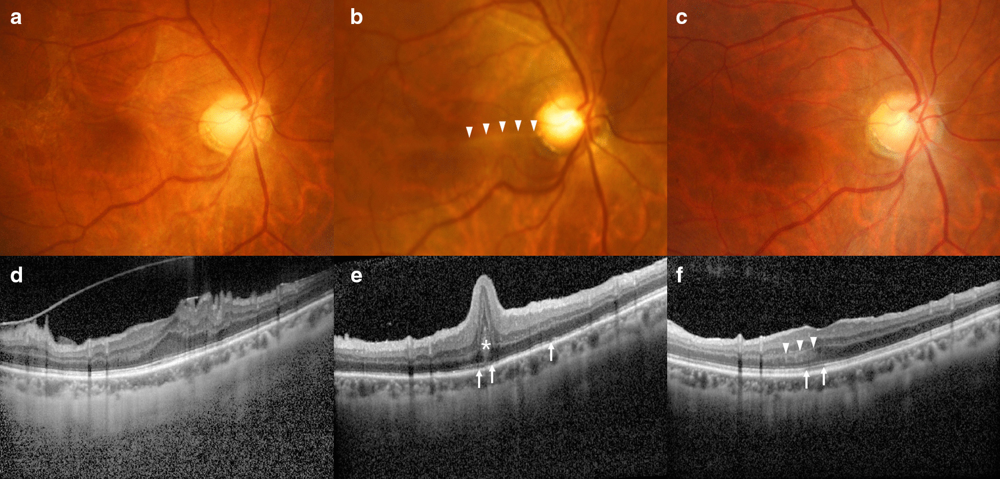
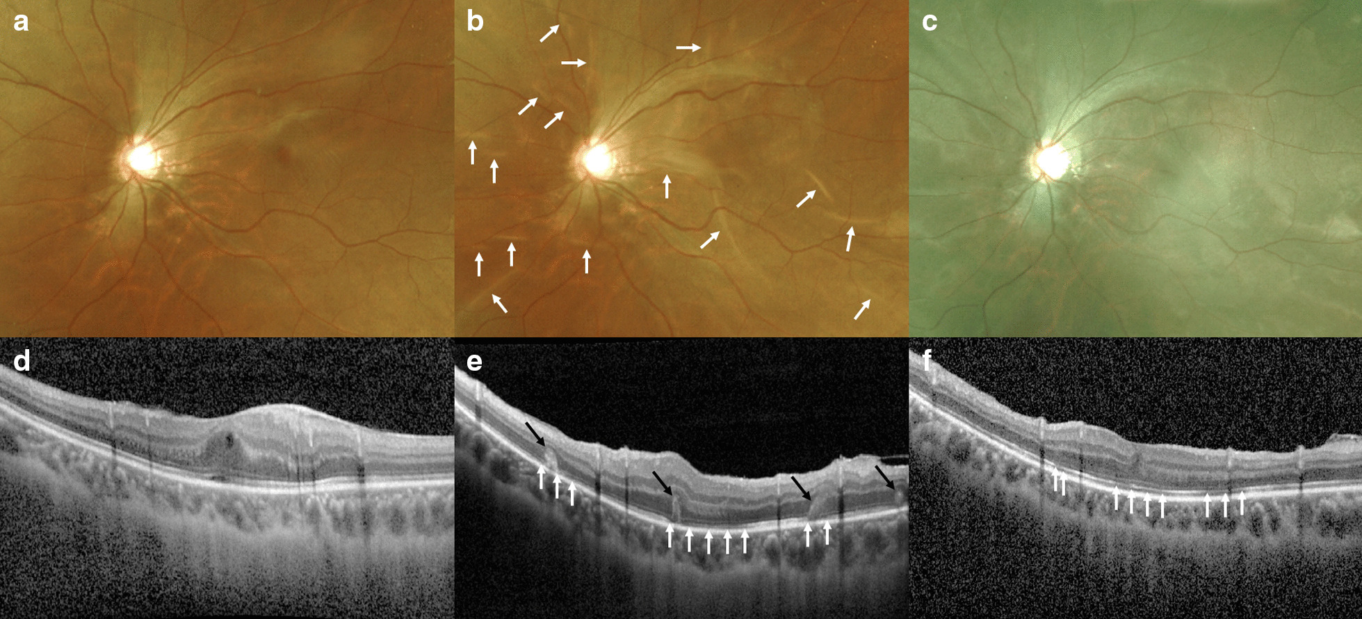
A 53-year-old Korean woman (case 1) underwent phacoemulsification and posterior chamber lens implantation combined with 25-gauge PPV for ERM removal in the right eye. On the following day, she had severe ocular hypotony, with an intraocular pressure (IOP) that was unmeasurable using a pneumatic tonometer. Despite normalization of IOP, macular retinal and photoreceptor folds with photoreceptor disruptions developed, and Henle's fiber layer hyperreflectivity was identified. Thereafter, retinal and photoreceptor folds gradually disappeared but photoreceptor disruption and Henle's fiber layer hyperreflectivity did not improve until 1 year postoperatively, with persistent central visual field distortion and visual acuity worse than that at the preoperative state. A 20-year-old Korean man (case 2) underwent an additional 25-gauge PPV for ERM removal in the left eye. Examination on the following day showed ocular hypotony and retinal folds with peripheral choroidal detachment. Although IOP was normalized, further OCT revealed photoreceptor folds and photoreceptor disruptions. Since then, the photoreceptor folds resolved; however, the photoreceptor disruption remained in the macula at the 1-year follow up, with persistent distorted vision and visual acuity worse than that at the preoperative state.
Early hypotony after vitrectomy for ERM could result in maculopathy leading to irreversible visual decline and metamorphopsia. Photoreceptor folds on OCT are characteristic features and the predominant mechanism of central visual loss in cases of hypotony maculopathy.
低眼压性黄斑病变通常被认为是青光眼手术或眼外伤的并发症。仅有少数关于玻璃体切割术后(PPV)发生低眼压性黄斑病变的报道。在此,我们报告两例因特发性黄斑裂孔而接受经睫状体平坦部玻璃体切除术(PPV)治疗的病例,并观察到光学相干断层扫描(OCT)下特征性的光感受器折叠。
一名 53 岁韩国女性(病例 1)右眼行超声乳化白内障吸除术和后房型人工晶状体植入术联合 25G PPV 治疗特发性黄斑裂孔。次日,患者出现严重眼球低眼压,气动眼压计无法测量眼压。尽管眼压恢复正常,但出现黄斑视网膜和光感受器折叠,伴有光感受器破坏,Henle 纤维层高反射。此后,视网膜和光感受器折叠逐渐消失,但光感受器破坏和 Henle 纤维层高反射直到术后 1 年仍未改善,且中心视野扭曲和视力较术前差。一名 20 岁韩国男性(病例 2)左眼行额外的 25G PPV 治疗特发性黄斑裂孔。次日检查发现眼球低眼压和视网膜折叠伴周边脉络膜脱离。尽管眼压恢复正常,但进一步的 OCT 显示光感受器折叠和光感受器破坏。此后,光感受器折叠消退;然而,在 1 年的随访中,黄斑区仍存在光感受器破坏,视力持续扭曲,视力较术前差。
ERM 玻璃体切除术后早期低眼压可能导致黄斑病变,导致不可逆转的视力下降和视物变形。OCT 下的光感受器折叠是低眼压性黄斑病变导致中心视力丧失的特征性表现和主要机制。