Reinert Dominik, Mönnings Peter, Schneider Ruth, Lukas Carsten
Department of Radiology, St. Josef Hospital Ruhr University Bochum, Bochum, Germany.
Department of Neurology, St. Josef Hospital Ruhr University Bochum, Bochum, Germany.
Radiol Case Rep. 2021 Jul;16(7):1815-1818. doi: 10.1016/j.radcr.2021.04.047. Epub 2021 Apr 30.
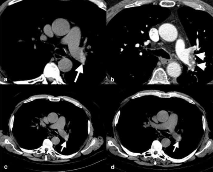
The request for CT (computed tomography) diagnostic in patients with suspected COVID-19 pneumonia has become part of the daily clinical routine. We reported a case of a 61-year-old patient with flu-like symptoms and a suspected COVID-19 pneumonia. After a negative PCR-test (polymerase chain reaction), a non-contrast enhanced CT was performed which revealed a suspicious hyperdensity in the left pulmonary artery and a pneumonia in the left lower lobe. A contrast enhanced CT confirmed a pulmonary embolism. An acute pulmonary embolism is a major complication and a main differential diagnosis of COVID-19. A hyperdense pulmonary artery sign (PAS) is a sensitive sign for a pulmonary embolism. Non-enhanced chest CT scans should be checked for hyperdense PAS in suspected of COVID-19 patients.
对疑似新型冠状病毒肺炎患者进行CT(计算机断层扫描)诊断的要求已成为日常临床工作的一部分。我们报告了一例61岁有流感样症状且疑似新型冠状病毒肺炎的患者。在聚合酶链反应(PCR)检测呈阴性后,进行了非增强CT检查,结果显示左肺动脉有可疑高密度影以及左下叶肺炎。增强CT证实为肺栓塞。急性肺栓塞是新型冠状病毒肺炎的主要并发症和主要鉴别诊断之一。肺动脉高密度征(PAS)是肺栓塞的一个敏感征象。对于疑似新型冠状病毒肺炎患者,应在非增强胸部CT扫描中检查是否存在肺动脉高密度征。