• 文献检索
  • 文档翻译
  • 深度研究
  • 学术资讯
  • Suppr Zotero 插件Zotero 插件
  • 邀请有礼
  • 套餐&价格
  • 历史记录
应用&插件
Suppr Zotero 插件Zotero 插件浏览器插件Mac 客户端Windows 客户端微信小程序
定价
高级版会员购买积分包购买API积分包
服务
文献检索文档翻译深度研究API 文档MCP 服务
关于我们
关于 Suppr公司介绍联系我们用户协议隐私条款
关注我们

Suppr 超能文献

核心技术专利:CN118964589B侵权必究
粤ICP备2023148730 号-1Suppr @ 2026

文献检索

告别复杂PubMed语法,用中文像聊天一样搜索,搜遍4000万医学文献。AI智能推荐,让科研检索更轻松。

立即免费搜索

文件翻译

保留排版,准确专业,支持PDF/Word/PPT等文件格式,支持 12+语言互译。

免费翻译文档

深度研究

AI帮你快速写综述,25分钟生成高质量综述,智能提取关键信息,辅助科研写作。

立即免费体验

评估非增强计算机断层扫描在肺栓塞诊断中的性能。

Evaluating the Performance of Unenhanced Computed Tomography in the Diagnosis of Pulmonary Embolism.

作者信息

Ehsanbakhsh Alireza, Hatami Farbod, Valizadeh Niloufar, Khorashadizadeh Nasrin, Norouzirad Farshad

机构信息

Department of Radiology, Birjand University of Medical Sciences, Birjand, Iran.

Cardiovascular Diseases Research Center, Birjand University of Medical Sciences, Birjand, Iran.

出版信息

J Tehran Heart Cent. 2021 Oct;16(4):156-161. doi: 10.18502/jthc.v16i4.8601.

DOI:10.18502/jthc.v16i4.8601
PMID:35935550
原文链接:https://pmc.ncbi.nlm.nih.gov/articles/PMC9308882/
Abstract

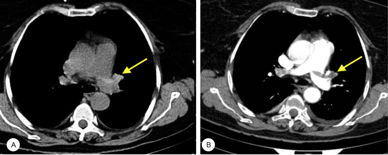
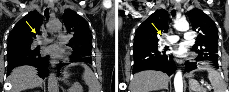
Computed tomography pulmonary angiography (CTPA) as the gold-standard examination in the detection of pulmonary embolism (PE) is contraindicated or unavailable in certain cases. The current study aimed to assess the accuracy of unenhanced CT in the diagnosis of PE. This cohort study was conducted between October 2020 and March 2021 in Birjand, Iran, on 195 participants with clinical suspicion of PE examined with multidetector computed tomography (MDCT) scanning and CTPA. The patients were categorized into 2 groups based on the diagnosis PE in CTPA results. Imaging variables in unenhanced CT scans, including hyper/hypodense intraluminal signs, pulmonary trunk enlargements, peripheral wedge-shaped opacities, and pleural effusions, were independently reviewed by 2 radiologists and then compared between the groups. There were 82 men (42.1%) and 113 women (57.9%) at a mean age ± standard deviation of 56.00±0.24 years. Based on CTPA results, PE was diagnosed in 24.1% of the study population (47/195). However, only 20 cases (42.5%) were detected by MDCT: 17 cases (85.0%) with central PE and 3 cases (15.0%) with peripheral PE. Concerning the intraluminal clot density, 12 patients (60.0%) had hyperdense signs, 3 (15.0%) had hypodense signs, and 5 (25.0%) had mixed hyper/hypodense signs. There was a significant difference between central PE and peripheral PE detected by MDCT. Intraluminal signs had the highest specificity and sensitivity (98.6% and 42.5%, area under the curve =0.734). Unenhanced MDCT has a remarkable performance in detecting PE, specifically central clots, and can, therefore, be considered an alternative modality when CTPA is not available or indicated.

摘要

计算机断层扫描肺动脉造影(CTPA)作为检测肺栓塞(PE)的金标准检查,在某些情况下是禁忌的或无法进行的。本研究旨在评估平扫CT诊断PE的准确性。这项队列研究于2020年10月至2021年3月在伊朗比尔詹德对195名临床怀疑患有PE的参与者进行,这些参与者接受了多排螺旋计算机断层扫描(MDCT)和CTPA检查。根据CTPA结果将患者分为两组。两名放射科医生独立评估平扫CT扫描中的影像变量,包括管腔内高密度/低密度征象、肺动脉主干增粗、外周楔形致密影和胸腔积液,然后在两组之间进行比较。共有82名男性(42.1%)和113名女性(57.9%),平均年龄±标准差为56.00±0.24岁。根据CTPA结果,24.1%的研究人群(47/195)被诊断为PE。然而,MDCT仅检测到20例(42.5%):17例(85.0%)为中央型PE,3例(15.0%)为外周型PE。关于管腔内血栓密度,12例患者(60.0%)有高密度征象,3例(15.0%)有低密度征象,5例(25.0%)有混合高密度/低密度征象。MDCT检测到的中央型PE和外周型PE之间存在显著差异。管腔内征象具有最高的特异性和敏感性(98.6%和42.5%,曲线下面积=0.734)。平扫MDCT在检测PE方面,特别是中央型血栓方面具有显著性能,因此,当无法进行或不适合CTPA时,可将其视为一种替代检查方法。

https://cdn.ncbi.nlm.nih.gov/pmc/blobs/4e02/9308882/0a2b4f343f41/JTHC-16-156-g002.jpg
https://cdn.ncbi.nlm.nih.gov/pmc/blobs/4e02/9308882/5e797cd01d01/JTHC-16-156-g001.jpg
https://cdn.ncbi.nlm.nih.gov/pmc/blobs/4e02/9308882/0a2b4f343f41/JTHC-16-156-g002.jpg
https://cdn.ncbi.nlm.nih.gov/pmc/blobs/4e02/9308882/5e797cd01d01/JTHC-16-156-g001.jpg
https://cdn.ncbi.nlm.nih.gov/pmc/blobs/4e02/9308882/0a2b4f343f41/JTHC-16-156-g002.jpg

相似文献

1
Evaluating the Performance of Unenhanced Computed Tomography in the Diagnosis of Pulmonary Embolism.评估非增强计算机断层扫描在肺栓塞诊断中的性能。
J Tehran Heart Cent. 2021 Oct;16(4):156-161. doi: 10.18502/jthc.v16i4.8601.
2
Unenhanced multidetector computed tomography findings in acute central pulmonary embolism.急性中央型肺栓塞的多排 CT 平扫表现。
BMC Med Imaging. 2019 Aug 14;19(1):65. doi: 10.1186/s12880-019-0364-y.
3
Diagnostic accuracy of three-dimensional contrast-enhanced MR angiography at 3-T for acute pulmonary embolism detection: comparison with multidetector CT angiography.3-T场强下三维对比增强磁共振血管造影对急性肺栓塞检测的诊断准确性:与多排CT血管造影的比较
Int J Cardiol. 2013 Oct 12;168(5):4775-83. doi: 10.1016/j.ijcard.2013.07.228. Epub 2013 Aug 1.
4
Parenchymal and pleural abnormalities in children with and without pulmonary embolism at MDCT pulmonary angiography.多层螺旋 CT 肺动脉造影中肺栓塞患儿与非肺栓塞患儿的实质和胸膜异常。
Pediatr Radiol. 2010 Feb;40(2):173-81. doi: 10.1007/s00247-009-1418-z. Epub 2009 Oct 22.
5
Multislice computed tomography angiography as an imaging modality of choice in patients with suspicion of pulmonary embolism - own experiences and modern imaging techniques.多层螺旋 CT 血管造影作为疑似肺栓塞患者的首选成像方式 - 自身经验和现代成像技术。
Adv Clin Exp Med. 2013 Sep-Oct;22(5):705-13.
6
Intraindividual comparison of gadolinium- and iodine-enhanced 64-slice multidetector CT pulmonary angiography for the detection of pulmonary embolism in a porcine model.钆增强与碘增强64层多排螺旋CT肺血管造影术在猪模型中检测肺栓塞的个体内比较
Emerg Radiol. 2011 Jun;18(3):189-95. doi: 10.1007/s10140-010-0926-3. Epub 2010 Dec 7.
7
Sensitivity and specificity of chest ultrasound in the diagnosis of pulmonary embolism in the emergency department: A case-control study.急诊科胸部超声诊断肺栓塞的敏感性和特异性:一项病例对照研究。
Can J Respir Ther. 2023 Jun 6;59:123-129. doi: 10.29390/cjrt-2022-065. eCollection 2023.
8
Can we rely on computed tomographic scanning to diagnose pulmonary embolism in critically ill surgical patients?我们能否依靠计算机断层扫描来诊断重症外科患者的肺栓塞?
J Trauma. 2004 Mar;56(3):518-25; discussion 525-6. doi: 10.1097/01.ta.0000114535.64175.c5.
9
Imaging modalities for confirming pulmonary embolism during pregnancy: results from a multicenter international study.孕期确诊肺栓塞的影像学检查方法:一项多中心国际研究的结果
Eur Radiol. 2022 Feb;32(2):1238-1246. doi: 10.1007/s00330-021-08161-9. Epub 2021 Jul 30.
10
Gallium-68 Ventilation/Perfusion PET-CT and CT Pulmonary Angiography for Pulmonary Embolism Diagnosis: An Interobserver Agreement Study.镓-68通气/灌注正电子发射断层显像X线计算机体层摄影术与CT肺血管造影术用于肺栓塞诊断:一项观察者间一致性研究
Front Med (Lausanne). 2021 Feb 16;7:599901. doi: 10.3389/fmed.2020.599901. eCollection 2020.

引用本文的文献

1
Usefulness of electron density imaging from dual-energy CT for detecting pulmonary thromboembolism in unenhanced CT.双能CT电子密度成像在未增强CT中检测肺血栓栓塞症的应用价值
Eur Radiol. 2025 Jul 17. doi: 10.1007/s00330-025-11846-0.
2
Advancements in Machine Learning and Artificial Intelligence in the Radiological Detection of Pulmonary Embolism.机器学习与人工智能在肺栓塞放射学检测中的进展
Cureus. 2025 Jan 29;17(1):e78217. doi: 10.7759/cureus.78217. eCollection 2025 Jan.
3
Chest non‑contrasted computed tomography in detecting acute pulmonary thromboembolism: A single‑center retrospective study.

本文引用的文献

1
Hyperdense pulmonary artery sign - detection of pulmonary embolism in patients with suspected COVID-19 using non-contrast chest CT.高密度肺动脉征——使用非增强胸部CT对疑似新型冠状病毒肺炎患者进行肺栓塞检测
Radiol Case Rep. 2021 Jul;16(7):1815-1818. doi: 10.1016/j.radcr.2021.04.047. Epub 2021 Apr 30.
2
Pulmonary embolism: update on management and controversies.肺栓塞:管理与争议的更新。
BMJ. 2020 Aug 5;370:m2177. doi: 10.1136/bmj.m2177.
3
Unenhanced multidetector computed tomography findings in acute central pulmonary embolism.
胸部非增强计算机断层扫描在检测急性肺血栓栓塞症中的应用:一项单中心回顾性研究。
Exp Ther Med. 2024 May 29;28(2):304. doi: 10.3892/etm.2024.12593. eCollection 2024 Aug.
4
Thrombus discrimination with electron density: potential implications for non-contrast computed tomography imaging.基于电子密度的血栓鉴别:对非增强计算机断层扫描成像的潜在影响。
Cardiovasc Diagn Ther. 2024 Apr 30;14(2):304-310. doi: 10.21037/cdt-24-28. Epub 2024 Apr 22.
5
Development and validation of a lung graph-based machine learning model to predict acute pulmonary thromboembolism on chest noncontrast computed tomography.基于肺部影像的机器学习模型的开发与验证,用于在胸部非增强计算机断层扫描上预测急性肺血栓栓塞症。
Quant Imaging Med Surg. 2023 Oct 1;13(10):6710-6723. doi: 10.21037/qims-22-1059. Epub 2023 Sep 1.
急性中央型肺栓塞的多排 CT 平扫表现。
BMC Med Imaging. 2019 Aug 14;19(1):65. doi: 10.1186/s12880-019-0364-y.
4
Epidemiology, Pathophysiology, and Natural History of Pulmonary Embolism.肺栓塞的流行病学、病理生理学及自然史
Semin Intervent Radiol. 2018 Jun;35(2):92-98. doi: 10.1055/s-0038-1642036. Epub 2018 Jun 4.
5
The validity of hyperdense lumen sign in non-contrast chest CT scans in the detection of pulmonary thromboembolism.非增强胸部 CT 扫描中高密度管腔征对肺血栓栓塞症的检测价值。
Int J Cardiovasc Imaging. 2011 Mar;27(3):433-40. doi: 10.1007/s10554-010-9673-5. Epub 2010 Jul 24.
6
Clinical usefulness of computed tomography study without contrast injection in the evaluation of acute pulmonary embolism.非增强计算机断层扫描在急性肺栓塞评估中的临床应用价值
J Comput Assist Tomogr. 2005 Jan-Feb;29(1):6-12. doi: 10.1097/01.rct.0000148274.45419.95.
7
Visualization of hypoattenuation clots on unenhanced CT of the thorax.
AJR Am J Roentgenol. 2004 Feb;182(2):530-1. doi: 10.2214/ajr.182.2.1820530.
8
Prediction of anemia on unenhanced computed tomography of the thorax.胸部平扫计算机断层扫描对贫血的预测
Can Assoc Radiol J. 2003 Feb;54(1):26-30.
9
Can computed tomography identify patients with anaemia?计算机断层扫描能识别贫血患者吗?
Ulster Med J. 2001 Nov;70(2):116-8.
10
Peripheral pulmonary arteries: how far in the lung does multi-detector row spiral CT allow analysis?
Radiology. 2001 Jun;219(3):629-36. doi: 10.1148/radiology.219.3.r01jn32629.