Liu Yujia, Fan Huijian, Dong Di, Liu Ping, He Bingxi, Meng Lingwei, Chen Jiaming, Chen Chunlin, Lang Jinghe, Tian Jie
School of Artificial Intelligence, University of Chinese Academy of Sciences, Beijing 100049, China; CAS Key Laboratory of Molecular Imaging, Institute of Automation, Chinese Academy of Sciences, Beijing 100190, China.
Department of Obstetrics and Gynecology, Nanfang Hospital, Southern Medical University, Guangzhou 510515, China.
Transl Oncol. 2021 Aug;14(8):101113. doi: 10.1016/j.tranon.2021.101113. Epub 2021 May 8.
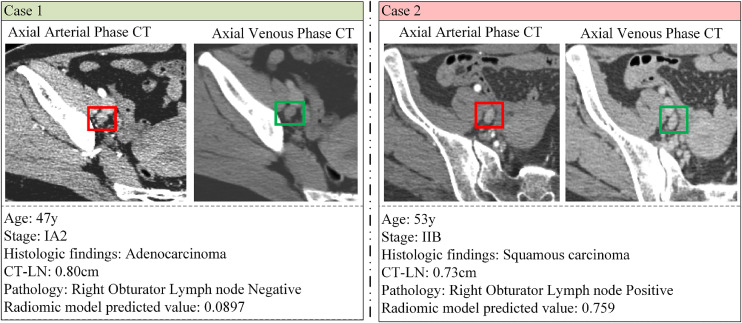
Radiomic models have been demonstrated to have acceptable discrimination capability for detecting lymph node metastasis (LNM). We aimed to develop a computed tomography-based radiomic model and validate its usefulness in the prediction of normal-sized LNM at node level in cervical cancer.
A total of 273 LNs of 219 patients from 10 centers were evaluated in this study. We randomly divided the LNs from the 2 centers with the largest number of LNs into the training and internal validation cohorts, and the rest as the external validation cohort. Radiomic features were extracted from the arterial and venous phase images. We trained an artificial neural network (ANN) to develop two single-phase models. A radiomic model reflecting the features of two-phase images was also built for directly predicting LNM in cervical cancer. Moreover, four state-of-the-art methods were used for comparison. The performance of all models was assessed using the area under the receiver operating characteristic curve (AUC).
Among the models we built, the models combining the features of two phases surpassed the single-phase models, and the models generated by ANN had better performance than the others. We found that the radiomic model achieved the highest AUCs of 0.912 and 0.859 in the training and internal validation cohorts, respectively. In the external validation cohort, the AUC of the radiomic model was 0.800.
We constructed a radiomic model that exhibited great ability in the prediction of LNM. The application of the model could optimize clinical staging and decision-making.
已证明放射组学模型在检测淋巴结转移(LNM)方面具有可接受的鉴别能力。我们旨在开发一种基于计算机断层扫描的放射组学模型,并验证其在预测宫颈癌中正常大小淋巴结水平的LNM方面的有用性。
本研究评估了来自10个中心的219例患者的273个淋巴结。我们将来自淋巴结数量最多的2个中心的淋巴结随机分为训练组和内部验证组,其余作为外部验证组。从动脉期和静脉期图像中提取放射组学特征。我们训练了一个人工神经网络(ANN)来开发两个单相模型。还建立了一个反映双期图像特征的放射组学模型,用于直接预测宫颈癌中的LNM。此外,使用了四种最先进的方法进行比较。所有模型的性能均使用受试者工作特征曲线下面积(AUC)进行评估。
在我们建立的模型中,结合双期特征的模型优于单相模型,ANN生成的模型性能优于其他模型。我们发现,放射组学模型在训练组和内部验证组中的AUC分别达到了最高的0.912和0.859。在外部验证组中,放射组学模型的AUC为0.800。
我们构建了一个在预测LNM方面表现出强大能力的放射组学模型。该模型的应用可以优化临床分期和决策。