Fan Yanfen, Yu Yixing, Wang Ximing, Hu Mengjie, Du Mingzhan, Guo Lingchuan, Sun Shifang, Hu Chunhong
Department of Radiology, The First Affiliated Hospital of Soochow University, Suzhou, Jiangsu, 215006, People's Republic of China.
Institute of Medical Imaging of Soochow University, Suzhou, Jiangsu, 215006, People's Republic of China.
J Hepatocell Carcinoma. 2021 May 5;8:349-359. doi: 10.2147/JHC.S293755. eCollection 2021.
To determine the potential findings associated with vessels encapsulating tumor clusters (VETC)-positive hepatocellular carcinoma (HCC), with particular emphasis on texture analysis based on gadolinium-ethoxybenzyl-diethylenetriamine pentaacetic acid (Gd-EOB-DTPA)-enhanced MRI.
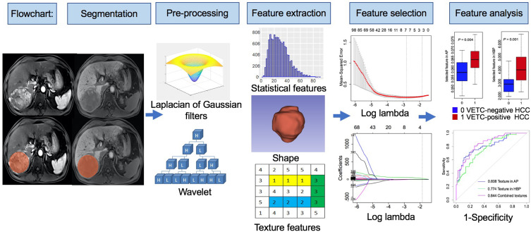
Eighty-one patients with VETC-negative HCC and 52 patients with VETC-positive HCC who underwent Gd-EOB-DTPA-enhanced MRI before curative partial hepatectomy were retrospectively evaluated in our institution. MRI texture analysis was performed on arterial phase (AP) and hepatobiliary phase (HBP) images. The least absolute shrinkage and selection operator (LASSO) logistic regression was used to select texture features most useful for identifying VETC-positive HCC. Univariate and multivariate analyses were used to determine significant variables for identifying the VETC-positive HCC in clinical factors and the texture features of MRI. Receiver operating characteristic (ROC) analysis and DeLong test were performed to compare the identified performances of significant variables for identifying VETC-positive HCC.
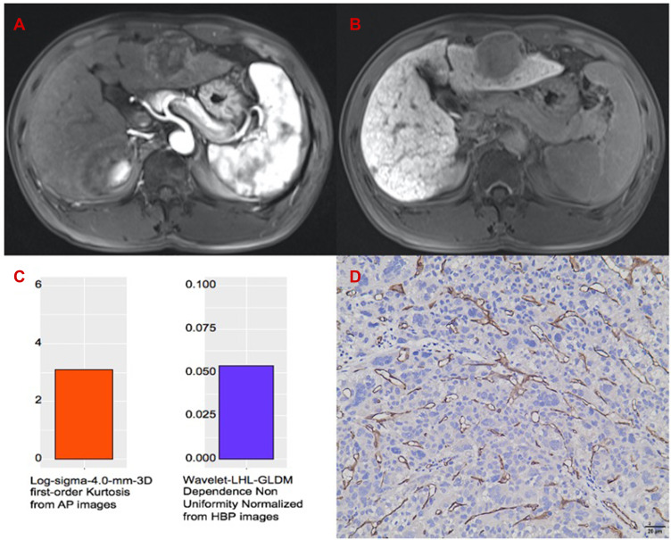
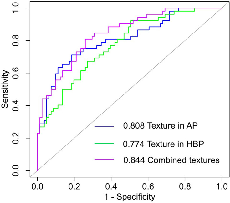
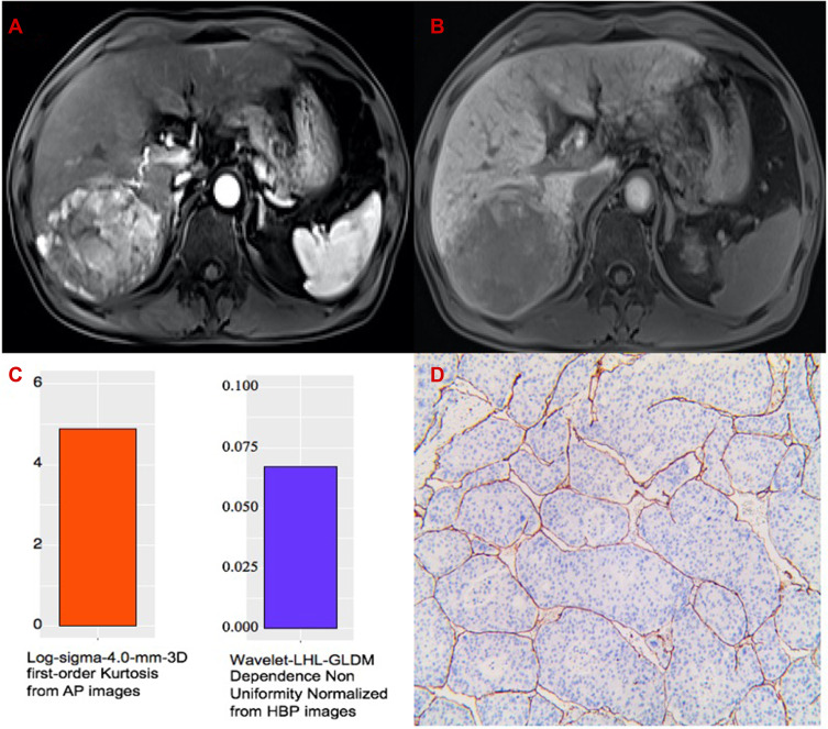
LASSO logistic regression selected 3 features in AP and HBP images, respectively. In multivariate analysis, the Log-sigma-4.0-mm-3D first-order Kurtosis derived from AP images (odds ratio [OR] = 4.128, = 0.001) and the Wavelet-LHL-GLDM Dependence Non Uniformity Normalized derived from HBP images (OR = 2.280, = 0.004) were independent significant variables associated with VETC-positive HCC. The combination of the two texture features for identifying VETC-positive HCC achieved an AUC value of 0.844 (95% confidence interval CI, 0.777, 0.910) with a sensitivity of 80.8% (95% CI, 70.1%, 91.5%) and specificity of 74.1% (95% CI, 64.5%, 83.6%).
Texture analysis based on Gd-EOB-DTPA-enhanced MRI can help identify VETC-positive HCC.
确定与包绕肿瘤细胞簇的血管(VETC)阳性肝细胞癌(HCC)相关的潜在发现,特别强调基于钆塞酸二钠(Gd-EOB-DTPA)增强磁共振成像(MRI)的纹理分析。
对我院81例VETC阴性HCC患者和52例VETC阳性HCC患者进行回顾性评估,这些患者在根治性部分肝切除术前均接受了Gd-EOB-DTPA增强MRI检查。对动脉期(AP)和肝胆期(HBP)图像进行MRI纹理分析。采用最小绝对收缩和选择算子(LASSO)逻辑回归来选择对识别VETC阳性HCC最有用的纹理特征。单因素和多因素分析用于确定临床因素和MRI纹理特征中识别VETC阳性HCC的显著变量。进行受试者操作特征(ROC)分析和DeLong检验,以比较识别VETC阳性HCC的显著变量的识别性能。
LASSO逻辑回归分别在AP和HBP图像中选择了3个特征。在多因素分析中,AP图像得出的Log-sigma-4.0-mm-3D一阶峰度(比值比[OR]=4.128,P=0.001)和HBP图像得出的小波-LHL-灰度共生矩阵依赖非均匀性归一化(OR=2.280,P=0.004)是与VETC阳性HCC相关的独立显著变量。这两种纹理特征联合识别VETC阳性HCC的曲线下面积(AUC)值为0.844(95%置信区间CI,0.777,0.910),灵敏度为80.8%(95%CI,70.1%,91.5%),特异度为74.1%(95%CI,64.5%,83.6%)。
基于Gd-EOB-DTPA增强MRI的纹理分析有助于识别VETC阳性HCC。