Matsuda Kosuke, Ueno Akihisa, Tsuzaki Junya, Kurebayashi Yutaka, Masugi Yohei, Yamazaki Ken, Tamura Masashi, Abe Yuta, Hasegawa Yasushi, Kitago Minoru, Jinzaki Masahiro, Sakamoto Michiie
Department of Pathology, Keio University School of Medicine, Tokyo, Japan.
Department of Pathology, Brigham and Women's Hospital, Harvard Medical School, Boston, Massachusetts, USA.
Hepatol Commun. 2024 Dec 11;9(1). doi: 10.1097/HC9.0000000000000593. eCollection 2025 Jan 1.
Vessels encapsulating tumor clusters (VETC) pattern is tumor vasculature of HCC and is a predictor of prognosis and therapeutic efficacy. Recent radiological studies have demonstrated the predictability of VETC from preoperative images, but the mechanisms of image formation are not elucidated. This study aims to determine the relationship between VETC and intratumor heterogeneity in Gd-EOB-DTPA-enhanced magnetic resonance imaging (EOB-MRI) and to provide its pathological evidence.
Radiologists visually classified preoperative arterial- and hepatobiliary-phase EOB-MRI images of 204 surgically resected HCCs into patterns based on heterogeneity and signal intensity; these classifications were validated using texture analysis. Single and multiplex immunohistochemistry for CD34, h-caldesmon, and OATP1B3 were performed to evaluate VETC, arterial vessel density (AVD), and OATP1B3 expression. Recurrence-free survival was assessed using the generalized Wilcoxon test. The contribution of clinicoradiological factors to the prediction of VETC was evaluated by random forest and least absolute shrinkage and selection operator regression.
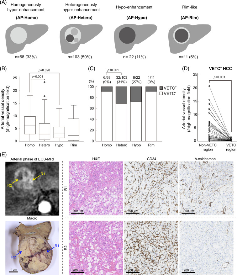
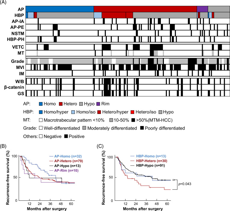
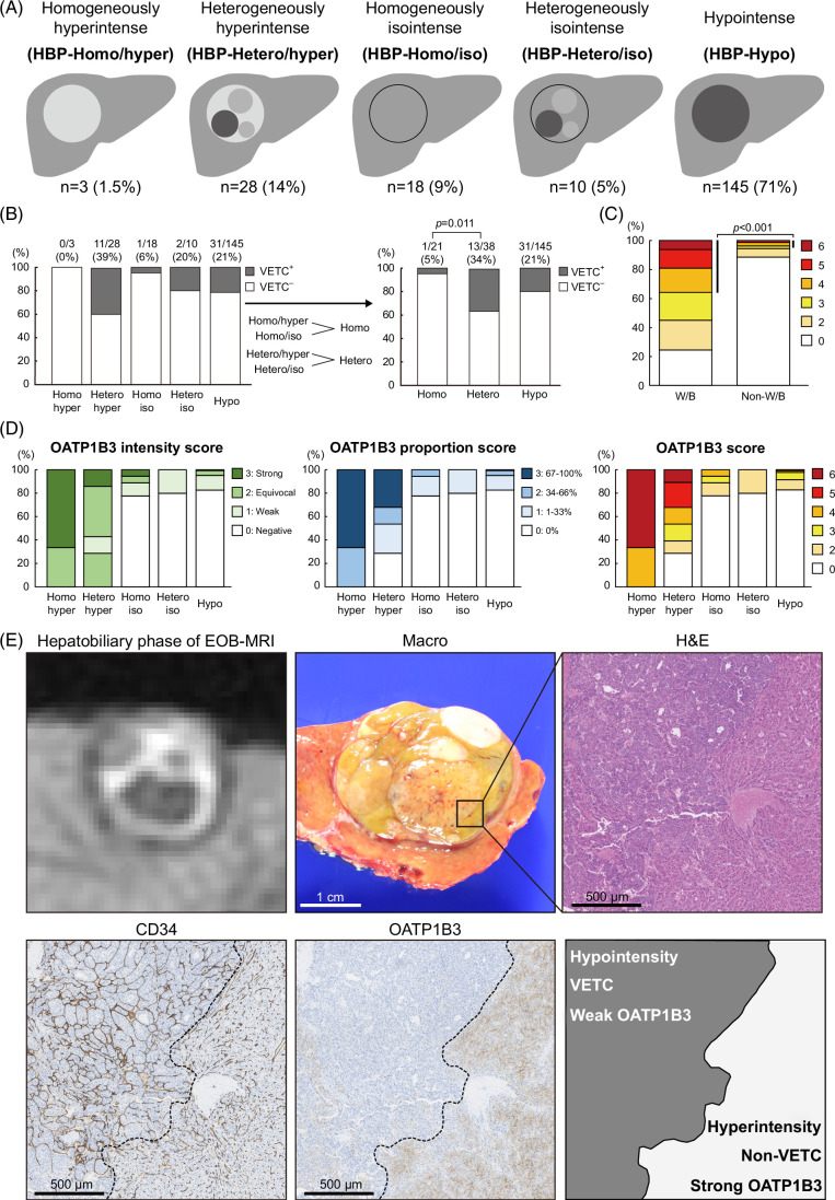
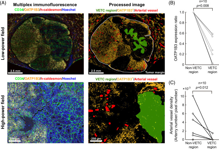
VETC was frequently found in tumors with arterial-phase heterogeneous hyper-enhancement patterns and in tumors with hepatobiliary-phase heterogeneous hyperintense/isointense patterns (HBP-Hetero). AVD and OATP1B3 expression positively correlated with signal intensity in the arterial and hepatobiliary phases, respectively. Intratumor spatial analysis revealed that AVD and OATP1B3 expression were lower in VETC regions than in tumor regions without VETC. Patients with HBP-Hetero tumors had shorter recurrence-free survival. Machine learning models highlighted the importance of serum PIVKA-II, tumor size, and enhancement pattern of arterial and hepatobiliary phase for VETC prediction.
VETC is associated with local reductions of both AVD and OATP1B3 expression, likely contributing to heterogeneous enhancement patterns in EOB-MRI. Evaluation of the arterial and hepatobiliary phases of EOB-MRI would enhance the predictability of VETC.
包裹肿瘤细胞簇的血管(VETC)模式是肝细胞癌的肿瘤血管系统,是预后和治疗效果的预测指标。近期的放射学研究已证实术前影像对VETC具有预测性,但影像形成机制尚未阐明。本研究旨在确定VETC与钆塞酸二钠增强磁共振成像(EOB-MRI)中肿瘤内异质性之间的关系,并提供其病理证据。
放射科医生根据异质性和信号强度,将204例手术切除的肝细胞癌术前动脉期和肝胆期EOB-MRI图像直观地分类为不同模式;这些分类通过纹理分析进行验证。对CD34、h-钙调蛋白和OATP1B3进行单重和多重免疫组织化学检测,以评估VETC、动脉血管密度(AVD)和OATP1B3表达。使用广义Wilcoxon检验评估无复发生存率。通过随机森林以及最小绝对收缩和选择算子回归评估临床放射学因素对VETC预测的贡献。
VETC常见于动脉期异质性高增强模式的肿瘤以及肝胆期异质性高信号/等信号模式(HBP-Hetero)的肿瘤中。AVD和OATP1B3表达分别与动脉期和肝胆期的信号强度呈正相关。肿瘤内空间分析显示,VETC区域的AVD和OATP1B3表达低于无VETC的肿瘤区域。HBP-Hetero肿瘤患者的无复发生存期较短。机器学习模型强调了血清异常凝血酶原、肿瘤大小以及动脉期和肝胆期增强模式对VETC预测的重要性。
VETC与AVD和OATP1B3表达的局部降低有关,可能导致EOB-MRI中的异质性增强模式。评估EOB-MRI的动脉期和肝胆期将提高VETC的预测性。