• 文献检索
  • 文档翻译
  • 深度研究
  • 学术资讯
  • Suppr Zotero 插件Zotero 插件
  • 邀请有礼
  • 套餐&价格
  • 历史记录
应用&插件
Suppr Zotero 插件Zotero 插件浏览器插件Mac 客户端Windows 客户端微信小程序
定价
高级版会员购买积分包购买API积分包
服务
文献检索文档翻译深度研究API 文档MCP 服务
关于我们
关于 Suppr公司介绍联系我们用户协议隐私条款
关注我们

Suppr 超能文献

核心技术专利:CN118964589B侵权必究
粤ICP备2023148730 号-1Suppr @ 2026

文献检索

告别复杂PubMed语法,用中文像聊天一样搜索,搜遍4000万医学文献。AI智能推荐,让科研检索更轻松。

立即免费搜索

文件翻译

保留排版,准确专业,支持PDF/Word/PPT等文件格式,支持 12+语言互译。

免费翻译文档

深度研究

AI帮你快速写综述,25分钟生成高质量综述,智能提取关键信息,辅助科研写作。

立即免费体验

牛植入前胚胎的三维实时成像:一种用于体外受精胚胎评估的新方法。

Three-Dimensional Live Imaging of Bovine Preimplantation Embryos: A New Method for IVF Embryo Evaluation.

作者信息

Masuda Yasumitsu, Hasebe Ryo, Kuromi Yasushi, Kobayashi Masayoshi, Urataki Kanako, Hishinuma Mitsugu, Ohbayashi Tetsuya, Nishimura Ryo

机构信息

Department of Animal Science, Tottori Livestock Research Center, Tottori, Japan.

SCREEN Holdings Co., Ltd., Kyoto, Japan.

出版信息

Front Vet Sci. 2021 Apr 26;8:639249. doi: 10.3389/fvets.2021.639249. eCollection 2021.

DOI:10.3389/fvets.2021.639249
PMID:33981741
原文链接:https://pmc.ncbi.nlm.nih.gov/articles/PMC8107228/
Abstract

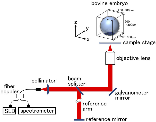
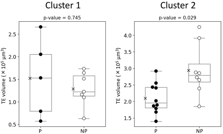
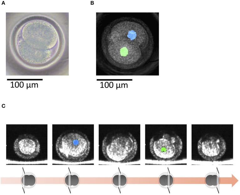
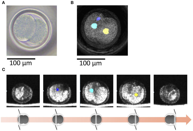
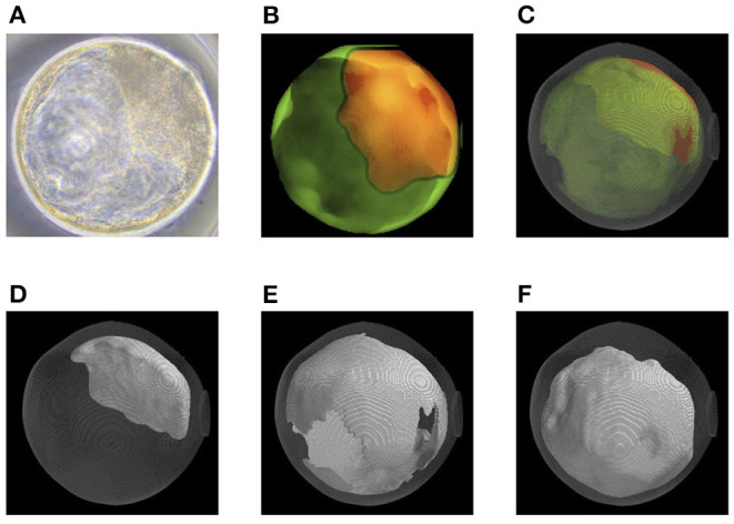
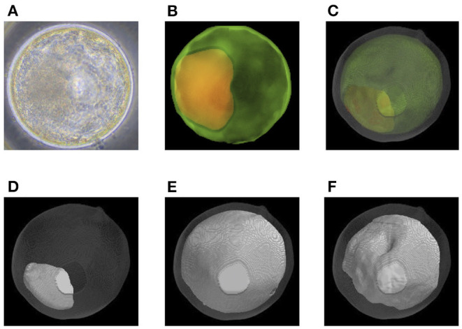
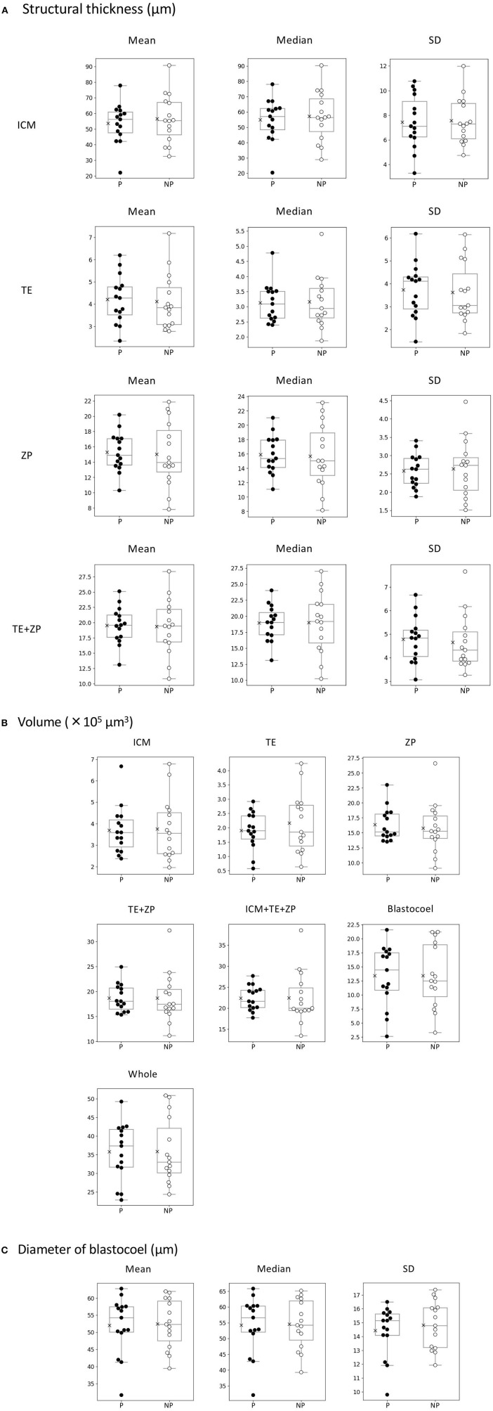
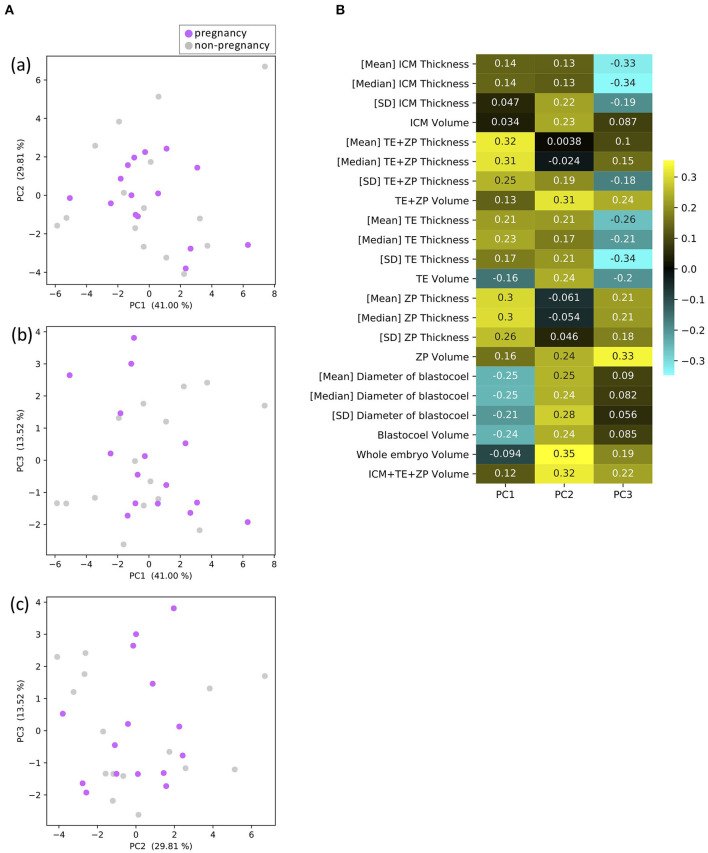
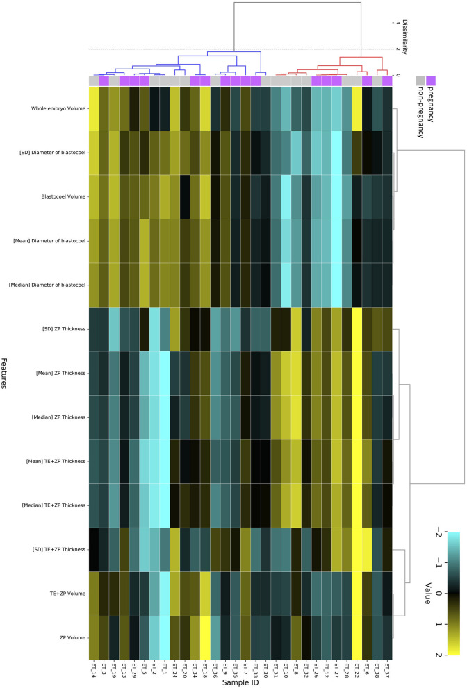
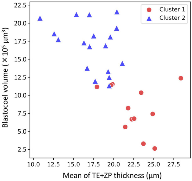
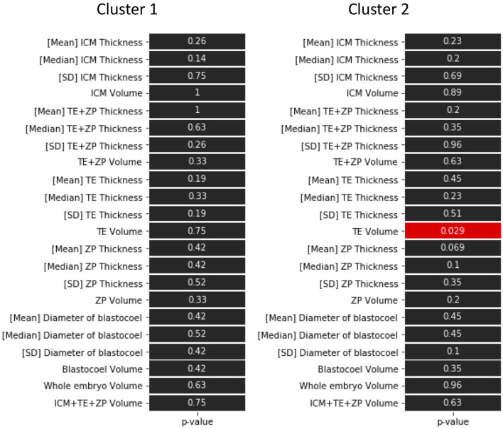
Conception rates for transferred bovine embryos are lower than those for artificial insemination. Embryo transfer (ET) is widely used in cattle but many of the transferred embryos fail to develop, thus, a more effective method for selecting bovine embryos suitable for ET is required. To evaluate the developmental potential of bovine preimplantation embryos (2-cell stage embryos and blastocysts), we have used the non-invasive method of optical coherence tomography (OCT) to obtain live images. The images were used to evaluate 22 parameters of blastocysts, such as the volume of the inner cell mass and the thicknesses of the trophectoderm (TE). Bovine embryos were obtained by fertilization (IVF) of the cumulus-oocyte complexes aspirated by ovum pick-up from Japanese Black cattle. The quality of the blastocysts was examined under an inverted microscope and all were confirmed to be Code1 according to the International Embryo Transfer Society standards for embryo evaluation. The OCT images of embryos were taken at the 2-cell and blastocyst stages prior to the transfer. In OCT, the embryos were irradiated with near-infrared light for a few minutes to capture three-dimensional images. Nuclei of the 2-cell stage embryos were clearly observed by OCT, and polynuclear cells at the 2-cell stage were also clearly found. With OCT, we were able to observe embryos at the blastocyst stage and evaluate their parameters. The conception rate following OCT (15/30; 50%) is typical for ETs and no newborn calves showed neonatal overgrowth or died, indicating that the OCT did not adversely affect the ET. A principal components analysis was unable to identify the parameters associated with successful pregnancy, while by using hierarchical clustering analysis, TE volume has been suggested to be one of the parameters for the evaluation of bovine embryo. The present results show that OCT imaging can be used to investigate time-dependent changes of IVF embryos. With further improvements, it should be useful for selecting high-quality embryos for transfer.

摘要

移植牛胚胎的受孕率低于人工授精的受孕率。胚胎移植(ET)在养牛业中被广泛应用,但许多移植的胚胎无法发育,因此,需要一种更有效的方法来选择适合胚胎移植的牛胚胎。为了评估牛植入前胚胎(2细胞期胚胎和囊胚)的发育潜力,我们使用了光学相干断层扫描(OCT)这种非侵入性方法来获取实时图像。这些图像用于评估囊胚的22个参数,如内细胞团的体积和滋养外胚层(TE)的厚度。通过从日本黑牛卵泡抽吸的卵丘-卵母细胞复合体进行体外受精(IVF)获得牛胚胎。在倒置显微镜下检查囊胚的质量,根据国际胚胎移植协会的胚胎评估标准,所有囊胚均被确认为1级。在移植前,在2细胞期和囊胚期拍摄胚胎的OCT图像。在OCT中,用近红外光照射胚胎几分钟以获取三维图像。通过OCT可以清晰地观察到2细胞期胚胎的细胞核,也能清楚地发现2细胞期的多核细胞。通过OCT,我们能够观察囊胚期的胚胎并评估其参数。OCT后的受孕率(15/30;50%)是胚胎移植的典型受孕率,没有新生犊牛出现新生儿过度生长或死亡的情况,这表明OCT对胚胎移植没有不利影响。主成分分析无法确定与成功妊娠相关的参数,而通过使用层次聚类分析,TE体积被认为是评估牛胚胎的参数之一。目前的结果表明,OCT成像可用于研究体外受精胚胎的时间依赖性变化。随着进一步改进,它应该有助于选择高质量的胚胎进行移植。

https://cdn.ncbi.nlm.nih.gov/pmc/blobs/a81d/8107228/7a0058dc38a3/fvets-08-639249-g0013.jpg
https://cdn.ncbi.nlm.nih.gov/pmc/blobs/a81d/8107228/27aff6513782/fvets-08-639249-g0001.jpg
https://cdn.ncbi.nlm.nih.gov/pmc/blobs/a81d/8107228/62fdd62e2d11/fvets-08-639249-g0002.jpg
https://cdn.ncbi.nlm.nih.gov/pmc/blobs/a81d/8107228/7afae5b2aa67/fvets-08-639249-g0003.jpg
https://cdn.ncbi.nlm.nih.gov/pmc/blobs/a81d/8107228/1e2172a92b74/fvets-08-639249-g0004.jpg
https://cdn.ncbi.nlm.nih.gov/pmc/blobs/a81d/8107228/c8e499d91637/fvets-08-639249-g0005.jpg
https://cdn.ncbi.nlm.nih.gov/pmc/blobs/a81d/8107228/6caac82126a7/fvets-08-639249-g0006.jpg
https://cdn.ncbi.nlm.nih.gov/pmc/blobs/a81d/8107228/50292aba197d/fvets-08-639249-g0007.jpg
https://cdn.ncbi.nlm.nih.gov/pmc/blobs/a81d/8107228/30fd12d906dd/fvets-08-639249-g0008.jpg
https://cdn.ncbi.nlm.nih.gov/pmc/blobs/a81d/8107228/2df75387b550/fvets-08-639249-g0009.jpg
https://cdn.ncbi.nlm.nih.gov/pmc/blobs/a81d/8107228/9d766e6a3110/fvets-08-639249-g0010.jpg
https://cdn.ncbi.nlm.nih.gov/pmc/blobs/a81d/8107228/b2ac1b02969c/fvets-08-639249-g0011.jpg
https://cdn.ncbi.nlm.nih.gov/pmc/blobs/a81d/8107228/32c88c35e951/fvets-08-639249-g0012.jpg
https://cdn.ncbi.nlm.nih.gov/pmc/blobs/a81d/8107228/7a0058dc38a3/fvets-08-639249-g0013.jpg

相似文献

1
Three-Dimensional Live Imaging of Bovine Preimplantation Embryos: A New Method for IVF Embryo Evaluation.牛植入前胚胎的三维实时成像:一种用于体外受精胚胎评估的新方法。
Front Vet Sci. 2021 Apr 26;8:639249. doi: 10.3389/fvets.2021.639249. eCollection 2021.
2
Hatchability evaluation of bovine IVF embryos using OCT-based 3D image analysis.使用基于 OCT 的 3D 图像分析评估牛体外受精胚胎的孵化能力。
J Reprod Dev. 2023 Oct 20;69(5):239-245. doi: 10.1262/jrd.2023-009. Epub 2023 Aug 11.
3
Three-dimensional live imaging of bovine embryos by optical coherence tomography.应用光学相干断层扫描技术对牛胚胎进行三维活体成像。
J Reprod Dev. 2021 Apr 21;67(2):149-154. doi: 10.1262/jrd.2020-151. Epub 2021 Jan 24.
4
Guidelines for the number of embryos to transfer following in vitro fertilization No. 182, September 2006.体外受精后胚胎移植数量指南,2006年9月,第182号
Int J Gynaecol Obstet. 2008 Aug;102(2):203-16. doi: 10.1016/j.ijgo.2008.01.007.
5
Three-dimensional morphology of bovine blastocysts hatched against lipopolysaccharide exposure in vitro.体外脂多糖暴露孵化的牛囊胚的三维形态。
Reprod Biol. 2024 Mar;24(1):100843. doi: 10.1016/j.repbio.2023.100843. Epub 2023 Dec 30.
6
Correlation between standard blastocyst morphology, euploidy and implantation: an observational study in two centers involving 956 screened blastocysts.标准囊胚形态、整倍体与着床的相关性:两个中心涉及 956 枚筛选囊胚的观察性研究。
Hum Reprod. 2014 Jun;29(6):1173-81. doi: 10.1093/humrep/deu033. Epub 2014 Feb 26.
7
Intrafollicular Oocyte Transfer (IFOT) of Abattoir-Derived and In Vitro-Matured Oocytes Results in Viable Blastocysts and Birth of Healthy Calves.屠宰场来源及体外成熟卵母细胞的卵泡内卵母细胞移植(IFOT)可产生有活力的囊胚并生出健康犊牛。
Biol Reprod. 2015 Jun;92(6):150. doi: 10.1095/biolreprod.114.124883. Epub 2015 Apr 29.
8
Offspring born from chimeras reconstructed from parthenogenetic and in vitro fertilized bovine embryos.由孤雌生殖和体外受精的牛胚胎重构而成的嵌合体所生育的后代。
Mol Reprod Dev. 1999 Jun;53(2):159-70. doi: 10.1002/(SICI)1098-2795(199906)53:2<159::AID-MRD5>3.0.CO;2-X.
9
Quality controls for bovine viral diarrhea virus-free IVF embryos.无牛病毒性腹泻病毒体外受精胚胎的质量控制
Theriogenology. 2000 Feb;53(3):827-39. doi: 10.1016/S0093-691X(99)00277-0.
10
What is the reproductive potential of day 7 euploid embryos?第 7 天的整倍体胚胎的生殖潜能如何?
Hum Reprod. 2019 Sep 29;34(9):1697-1706. doi: 10.1093/humrep/dez129.

引用本文的文献

1
Development of an AI-Assisted Embryo Selection System Using Iberian Ribbed Newts for Embryo-Fetal Development Toxicity Testing.开发一种基于伊比利亚肋突螈的人工智能辅助胚胎选择系统,用于胚胎-胎儿发育毒性测试。
Yonago Acta Med. 2024 Aug 27;67(3):233-241. doi: 10.33160/yam.2024.08.011. eCollection 2024 Aug.
2
Recent advancements to increase success in assisted reproductive technologies in cattle.提高奶牛辅助生殖技术成功率的最新进展。
Anim Reprod. 2024 Aug 12;21(3):e20240031. doi: 10.1590/1984-3143-AR2024-0031. eCollection 2024.
3
Excluding alanine from minimum essential medium (MEM) nonessential amino acid supplementation of the culture medium facilitates post-fertilization events and early cleavages of bovine oocytes fertilized in vitro.

本文引用的文献

1
Three-dimensional live imaging of bovine embryos by optical coherence tomography.应用光学相干断层扫描技术对牛胚胎进行三维活体成像。
J Reprod Dev. 2021 Apr 21;67(2):149-154. doi: 10.1262/jrd.2020-151. Epub 2021 Jan 24.
2
A 3D in vitro pericyte-supported microvessel model: visualisation and quantitative characterisation of multistep angiogenesis.一种3D体外周细胞支持的微血管模型:多步骤血管生成的可视化与定量表征
J Mater Chem B. 2018 Feb 21;6(7):1085-1094. doi: 10.1039/c7tb03239k. Epub 2018 Feb 5.
3
Direct transfer of frozen-thawed bovine embryos and its application in cattle reproduction management.
在体外受精的牛卵母细胞中,从最小必需培养基(MEM)非必需氨基酸补充物中去除丙氨酸有利于受精后的事件和早期卵裂。
J Reprod Dev. 2024 Aug 7;70(4):223-228. doi: 10.1262/jrd.2023-098. Epub 2024 May 17.
4
The number of nuclei in compacted embryos, assessed by optical coherence microscopy, is a non-invasive and robust marker of mouse embryo quality.利用光学相干显微镜评估致密胚胎中的细胞核数量是一种非侵入性的、强大的小鼠胚胎质量标志物。
Mol Hum Reprod. 2024 Mar 28;30(4). doi: 10.1093/molehr/gaae012.
5
Rapid Quantification of Microvessels of Three-Dimensional Blood-Brain Barrier Model Using Optical Coherence Tomography and Deep Learning Algorithm.使用光学相干断层扫描和深度学习算法快速定量三维血脑屏障模型中的微血管。
Biosensors (Basel). 2023 Aug 15;13(8):818. doi: 10.3390/bios13080818.
6
Hatchability evaluation of bovine IVF embryos using OCT-based 3D image analysis.使用基于 OCT 的 3D 图像分析评估牛体外受精胚胎的孵化能力。
J Reprod Dev. 2023 Oct 20;69(5):239-245. doi: 10.1262/jrd.2023-009. Epub 2023 Aug 11.
冻融牛胚胎的直接移植及其在奶牛繁殖管理中的应用。
J Reprod Dev. 2019 Oct 23;65(5):389-396. doi: 10.1262/jrd.2019-025. Epub 2019 Jun 13.
4
Live-cell imaging of nuclear-chromosomal dynamics in bovine in vitro fertilised embryos.牛体外受精胚胎中核染色体动力学的活细胞成像。
Sci Rep. 2018 May 10;8(1):7460. doi: 10.1038/s41598-018-25698-w.
5
Variations in bovine embryo production between individual donors for OPU-IVF are closely related to glutathione concentrations in oocytes during in vitro maturation.在OPU-IVF中,个体供体之间牛胚胎生产的差异与卵母细胞体外成熟过程中的谷胱甘肽浓度密切相关。
Theriogenology. 2018 Jun;113:176-182. doi: 10.1016/j.theriogenology.2018.03.002. Epub 2018 Mar 10.
6
A Vascular Endothelial Growth Factor-Dependent Sprouting Angiogenesis Assay Based on an In Vitro Human Blood Vessel Model for the Study of Anti-Angiogenic Drugs.基于体外人血管模型的血管内皮生长因子依赖性发芽血管生成分析法用于抗血管生成药物的研究。
EBioMedicine. 2018 Jan;27:225-236. doi: 10.1016/j.ebiom.2017.12.014. Epub 2017 Dec 20.
7
Speckle variance OCT for depth resolved assessment of the viability of bovine embryos.用于深度分辨评估牛胚胎活力的斑点方差光学相干断层扫描技术
Biomed Opt Express. 2017 Oct 20;8(11):5139-5150. doi: 10.1364/BOE.8.005139. eCollection 2017 Nov 1.
8
Genome stability of bovine in vivo-conceived cleavage-stage embryos is higher compared to in vitro-produced embryos.与体外受精产生的胚胎相比,体内受精的牛早期胚胎具有更高的基因组稳定性。
Hum Reprod. 2017 Nov 1;32(11):2348-2357. doi: 10.1093/humrep/dex286.
9
Optical coherence microscopy as a novel, non-invasive method for the 4D live imaging of early mammalian embryos.光学相干显微镜作为一种新颖的、非侵入性的方法,可用于对早期哺乳动物胚胎进行 4D 活体成像。
Sci Rep. 2017 Jun 23;7(1):4165. doi: 10.1038/s41598-017-04220-8.
10
Selection of viable in vitro-fertilized bovine embryos using time-lapse monitoring in microwell culture dishes.在微孔培养皿中使用延时监测技术筛选体外受精的 viable 牛胚胎。 (注:原文中“viable”可能有误,推测应为“viable”,意为“有活力的、能存活的” ,完整准确译文为:在微孔培养皿中使用延时监测技术筛选体外受精的有活力牛胚胎。 )
J Reprod Dev. 2017 Aug 19;63(4):353-357. doi: 10.1262/jrd.2017-041. Epub 2017 May 26.