• 文献检索
  • 文档翻译
  • 深度研究
  • 学术资讯
  • Suppr Zotero 插件Zotero 插件
  • 邀请有礼
  • 套餐&价格
  • 历史记录
应用&插件
Suppr Zotero 插件Zotero 插件浏览器插件Mac 客户端Windows 客户端微信小程序
定价
高级版会员购买积分包购买API积分包
服务
文献检索文档翻译深度研究API 文档MCP 服务
关于我们
关于 Suppr公司介绍联系我们用户协议隐私条款
关注我们

Suppr 超能文献

核心技术专利:CN118964589B侵权必究
粤ICP备2023148730 号-1Suppr @ 2026

文献检索

告别复杂PubMed语法,用中文像聊天一样搜索,搜遍4000万医学文献。AI智能推荐,让科研检索更轻松。

立即免费搜索

文件翻译

保留排版,准确专业,支持PDF/Word/PPT等文件格式,支持 12+语言互译。

免费翻译文档

深度研究

AI帮你快速写综述,25分钟生成高质量综述,智能提取关键信息,辅助科研写作。

立即免费体验

基于心脏 R2*采集的肝脏 R2*定量用于肝脏铁定量的可重复性。

Reproducibility of liver R2* quantification for liver iron quantification from cardiac R2* acquisitions.

机构信息

Department of Radiology, Wisconsin Institutes of Medical Research, University of Wisconsin, Room 2478, 1111 Highland Avenue, Madison, WI, 53705, USA.

Department of Radiology and Neuroradiology, University Greifswald, Greifswald, Germany.

出版信息

Abdom Radiol (NY). 2021 Sep;46(9):4200-4209. doi: 10.1007/s00261-021-03099-4. Epub 2021 May 12.

DOI:10.1007/s00261-021-03099-4
PMID:33982186
原文链接:https://pmc.ncbi.nlm.nih.gov/articles/PMC8346410/
Abstract

OBJECTIVES

To evaluate the reproducibility of liver R2* measurements between a 2D cardiac ECG-gated and a 3D breath-hold liver CSE-MRI acquisition for liver iron quantification.

METHODS

A total of 54 1.5 T MRI exams from 51 subjects (18 women, 36 men, age 35.2 ± 21.8) were included. These included two sub-studies with 23 clinical MRI exams from 19 patients identified retrospectively, 24 participants with known or suspected iron overload, and 7 healthy volunteers acquired prospectively. The 2D cardiac and the 3D liver R2* maps were acquired in the same exam. Either acquisitions were reconstructed using a complex R2* algorithm that accounts for the presence of fat and residual phase errors due to eddy currents. Data were analyzed using colocalized ROIs in the liver.

RESULTS

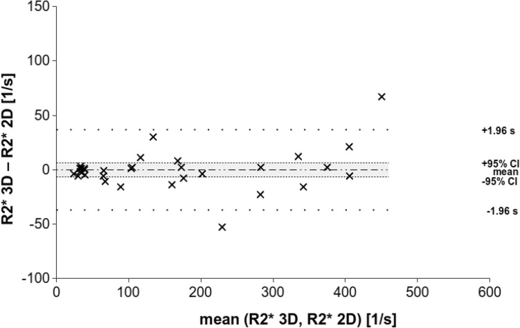
Linear regression analysis demonstrated high Pearson's correlation and Lin's concordance coefficient for the overall study and both sub-studies. Bland-Altman analysis also showed good agreement, except for a slight increase of the mean R2* value above ~ 400 s. The Kolmogorow-Smirnow test revealed a non-normal distribution for (R2* 3D-R2* 2D) values from 0 to 600 s in contrast to the 0-200 s and 0-400 s subpopulations. Linear regression analysis showed no relevant differences other than the intercept, likely due to only 7 measurements above 400 s.

CONCLUSIONS

The results demonstrate that R2*-measurements in the liver are feasible using 2D cardiac R2* maps compared to 3D liver R2* maps as the reference. Liver R2* may be underestimated for R2* > 400 s using the 2D cardiac R2* mapping method.

摘要

目的

评估二维心脏 ECG 门控和三维屏气肝脏 CSE-MRI 采集用于肝脏铁定量的肝脏 R2*测量的重现性。

方法

共纳入 51 例受试者(18 例女性,36 例男性,年龄 35.2±21.8 岁)的 54 例 1.5T MRI 检查。这些检查包括两项子研究,共 23 例临床 MRI 检查来自 19 例回顾性识别的患者,24 例已知或疑似铁过载参与者,以及 7 例健康志愿者前瞻性采集。在同一次检查中获得了二维心脏和三维肝脏 R2图谱。使用复杂的 R2算法重建两种采集,该算法考虑了脂肪的存在和由于涡流引起的残余相位误差。使用肝脏中的共定位 ROI 进行数据分析。

结果

线性回归分析表明,总体研究和两项子研究均显示出高 Pearson 相关系数和 Lin 一致性系数。Bland-Altman 分析也显示出良好的一致性,除了在~400s 以上的平均 R2值略有增加。Kolmogorow-Smirnow 检验显示,在 0 到 600s 的(R23D-R2*2D)值与 0 到 200s 和 0 到 400s 亚群的分布呈非正态分布。线性回归分析除了截距外,没有发现其他相关差异,可能是由于只有 7 次测量值大于 400s。

结论

结果表明,与使用 3D 肝脏 R2图谱作为参考相比,二维心脏 R2图谱可用于肝脏 R2测量。对于 R2>400s,使用二维心脏 R2映射方法可能会低估肝脏 R2。

https://cdn.ncbi.nlm.nih.gov/pmc/blobs/c7fb/8346410/904eb9328b30/261_2021_3099_Fig6_HTML.jpg
https://cdn.ncbi.nlm.nih.gov/pmc/blobs/c7fb/8346410/e51d761f20de/261_2021_3099_Fig1_HTML.jpg
https://cdn.ncbi.nlm.nih.gov/pmc/blobs/c7fb/8346410/82a57c215afd/261_2021_3099_Fig2_HTML.jpg
https://cdn.ncbi.nlm.nih.gov/pmc/blobs/c7fb/8346410/5295d1d89486/261_2021_3099_Fig3_HTML.jpg
https://cdn.ncbi.nlm.nih.gov/pmc/blobs/c7fb/8346410/ea652da79844/261_2021_3099_Fig4_HTML.jpg
https://cdn.ncbi.nlm.nih.gov/pmc/blobs/c7fb/8346410/df79f1b7a44c/261_2021_3099_Fig5_HTML.jpg
https://cdn.ncbi.nlm.nih.gov/pmc/blobs/c7fb/8346410/904eb9328b30/261_2021_3099_Fig6_HTML.jpg

相似文献

1
Reproducibility of liver R2* quantification for liver iron quantification from cardiac R2* acquisitions.基于心脏 R2*采集的肝脏 R2*定量用于肝脏铁定量的可重复性。
Abdom Radiol (NY). 2021 Sep;46(9):4200-4209. doi: 10.1007/s00261-021-03099-4. Epub 2021 May 12.
2
Hepatic Iron Quantification Using a Free-Breathing 3D Radial Gradient Echo Technique and Validation With a 2D Biopsy-Calibrated R Relaxometry Method.使用自由呼吸 3D 径向梯度回波技术进行肝脏铁定量及与 2D 活检校准 R 弛豫率法的验证。
J Magn Reson Imaging. 2022 May;55(5):1407-1416. doi: 10.1002/jmri.27921. Epub 2021 Sep 20.
3
Improved free-breathing liver fat and iron quantification using a 2D chemical shift-encoded MRI with flip angle modulation and motion-corrected averaging.使用具有翻转角调制和运动校正平均的二维化学位移编码 MRI 提高自由呼吸肝脏脂肪和铁定量的准确性。
Eur Radiol. 2022 Aug;32(8):5458-5467. doi: 10.1007/s00330-022-08682-x. Epub 2022 Mar 21.
4
Evaluation of liver iron overload with R2* relaxometry with versus without fat suppression: both are clinically accurate but there are differences.使用与不使用脂肪抑制的R2*弛豫测量法评估肝脏铁过载:两者在临床上均准确,但存在差异。
Eur Radiol. 2020 Nov;30(11):5826-5833. doi: 10.1007/s00330-020-07010-5. Epub 2020 Jun 13.
5
Comparison of Inline R2* MRI versus FerriScan for liver iron quantification in patients on chelation therapy for iron overload: preliminary results.比较在线 R2* MRI 与 FerriScan 在螯合治疗铁过载患者中肝脏铁定量的结果:初步结果。
Eur Radiol. 2021 Dec;31(12):9296-9305. doi: 10.1007/s00330-021-08019-0. Epub 2021 May 26.
6
Limits of Fat Quantification in the Presence of Iron Overload.铁过载状态下的脂肪定量的局限性。
J Magn Reson Imaging. 2021 Oct;54(4):1166-1174. doi: 10.1002/jmri.27611. Epub 2021 Mar 29.
7
Performance of different Dixon-based methods for MR liver iron assessment in comparison to a biopsy-validated R2* relaxometry method.不同基于 Dixon 方法的磁共振肝脏铁评估与经肝活检验证的 R2*弛豫率法的性能比较。
Eur Radiol. 2021 Apr;31(4):2252-2262. doi: 10.1007/s00330-020-07291-w. Epub 2020 Sep 23.
8
Complex confounder-corrected R2* mapping for liver iron quantification with MRI.采用复杂混杂因素校正 R2* 映射法对 MRI 肝脏铁定量。
Eur Radiol. 2021 Jan;31(1):264-275. doi: 10.1007/s00330-020-07123-x. Epub 2020 Aug 12.
9
Application of the magnetic resonance 3D multiecho Dixon sequence for quantifying hepatic iron overload and steatosis in patients with thalassemia.磁共振 3D 多回波 Dixon 序列在铁过载和脂肪变性定量分析地中海贫血患者肝脏中的应用。
Magn Reson Imaging. 2024 Sep;111:28-34. doi: 10.1016/j.mri.2024.03.015. Epub 2024 Mar 15.
10
Interexamination repeatability and spatial heterogeneity of liver iron and fat quantification using MRI-based multistep adaptive fitting algorithm.基于MRI的多步自适应拟合算法对肝脏铁和脂肪定量的检查间重复性及空间异质性
J Magn Reson Imaging. 2015 Nov;42(5):1281-90. doi: 10.1002/jmri.24922. Epub 2015 Apr 27.

引用本文的文献

1
Practical Application of Multivendor MRI-Based R2* Mapping for Liver Iron Quantification at 1.5 T and 3.0 T.基于多厂商磁共振成像的R2*映射在1.5T和3.0T肝脏铁定量中的实际应用
J Magn Reson Imaging. 2025 Jan;61(1):150-165. doi: 10.1002/jmri.29401. Epub 2024 Apr 25.
2
Quantifying Liver Heterogeneity via R2*-MRI with Super-Paramagnetic Iron Oxide Nanoparticles (SPION) to Characterize Liver Function and Tumor.通过使用超顺磁性氧化铁纳米颗粒(SPION)的R2*磁共振成像对肝脏异质性进行量化,以表征肝功能和肿瘤。
Cancers (Basel). 2022 Oct 27;14(21):5269. doi: 10.3390/cancers14215269.
3
Multicenter Reproducibility of Liver Iron Quantification with 1.5-T and 3.0-T MRI.

本文引用的文献

1
Complex confounder-corrected R2* mapping for liver iron quantification with MRI.采用复杂混杂因素校正 R2* 映射法对 MRI 肝脏铁定量。
Eur Radiol. 2021 Jan;31(1):264-275. doi: 10.1007/s00330-020-07123-x. Epub 2020 Aug 12.
2
Ensemble Learning for Early-Response Prediction of Antidepressant Treatment in Major Depressive Disorder.基于集成学习的重度抑郁症抗抑郁治疗早期反应预测。
J Magn Reson Imaging. 2020 Jul;52(1):161-171. doi: 10.1002/jmri.27029. Epub 2019 Dec 20.
3
Diversity in the medical workforce is better for patients.医疗劳动力的多样性对患者更有益。
1.5T 和 3.0T MRI 肝脏铁定量的多中心可重复性。
Radiology. 2023 Feb;306(2):e213256. doi: 10.1148/radiol.213256. Epub 2022 Oct 4.
4
Hepatic Iron Quantification Using a Free-Breathing 3D Radial Gradient Echo Technique and Validation With a 2D Biopsy-Calibrated R Relaxometry Method.使用自由呼吸 3D 径向梯度回波技术进行肝脏铁定量及与 2D 活检校准 R 弛豫率法的验证。
J Magn Reson Imaging. 2022 May;55(5):1407-1416. doi: 10.1002/jmri.27921. Epub 2021 Sep 20.
BMJ. 2019 Dec 11;367:l6874. doi: 10.1136/bmj.l6874.
4
Hyperferritinemia and inflammation.高血铁症与炎症。
Int Immunol. 2017 Nov 1;29(9):401-409. doi: 10.1093/intimm/dxx031.
5
Myocardial iron content and mitochondrial function in human heart failure: a direct tissue analysis.人心力衰竭中心肌铁含量与线粒体功能:直接的组织分析。
Eur J Heart Fail. 2017 Apr;19(4):522-530. doi: 10.1002/ejhf.640. Epub 2016 Sep 19.
6
MRI-based quantitative susceptibility mapping (QSM) and R2* mapping of liver iron overload: Comparison with SQUID-based biomagnetic liver susceptometry.基于磁共振成像的肝脏铁过载定量磁化率成像(QSM)和R2*成像:与基于超导量子干涉仪的生物磁肝脏磁测量法的比较
Magn Reson Med. 2017 Jul;78(1):264-270. doi: 10.1002/mrm.26358. Epub 2016 Aug 11.
7
Retrospective comparison of gradient recalled echo R2* and spin-echo R2 magnetic resonance analysis methods for estimating liver iron content in children and adolescents.梯度回波R2*与自旋回波R2磁共振分析方法用于评估儿童和青少年肝脏铁含量的回顾性比较
Pediatr Radiol. 2015 Oct;45(11):1629-34. doi: 10.1007/s00247-015-3378-9. Epub 2015 May 26.
8
Cardiac iron overload in sickle-cell disease.镰状细胞病中的心脏铁过载。
Am J Hematol. 2014 Jul;89(7):678-83. doi: 10.1002/ajh.23721. Epub 2014 Apr 15.
9
Quantification of liver iron with MRI: state of the art and remaining challenges.磁共振成像对肝脏铁含量的定量分析:现状与尚存的挑战
J Magn Reson Imaging. 2014 Nov;40(5):1003-21. doi: 10.1002/jmri.24584. Epub 2014 Mar 3.
10
Magnetic resonance imaging of the pediatric liver: imaging of steatosis, iron deposition, and fibrosis.小儿肝脏的磁共振成像:脂肪变性、铁沉积和纤维化的成像
Magn Reson Imaging Clin N Am. 2013 Nov;21(4):669-80. doi: 10.1016/j.mric.2013.05.001. Epub 2013 Aug 9.