Department of Radiology and Nuclear Medicine, Amsterdam University Medical Centres, Amsterdam, The Netherlands.
Department of Internal and Vascular Medicine, Amsterdam University Medical Centres, Amsterdam, The Netherlands.
J Magn Reson Imaging. 2021 Dec;54(6):1937-1949. doi: 10.1002/jmri.27703. Epub 2021 May 15.
Noninvasive diagnostic methods are urgently required in disease stratification and monitoring in nonalcoholic fatty liver disease (NAFLD). Multiparametric magnetic resonance imaging (MRI) is a promising technique to assess hepatic steatosis, inflammation, and fibrosis, potentially enabling noninvasive identification of individuals with active and advanced stages of NAFLD.
To examine the diagnostic performance of multiparametric MRI for the assessment of disease severity along the NAFLD disease spectrum with comparison to histological scores.
Prospective, cohort.
Thirty-seven patients with NAFLD.
FIELD STRENGTH/SEQUENCE: Multiparametric MRI at 3.0 T consisted of magnetic resonance (MR) spectroscopy (MRS) with multi-echo stimulated-echo acquisition mode, magnitude-based and three-point Dixon using a two-dimensional multi-echo gradient echo, MR elastography (MRE) using a generalized multishot gradient-recalled echo sequence and intravoxel incoherent motion (IVIM) using a multislice diffusion weighted single-shot echo-planar sequence.
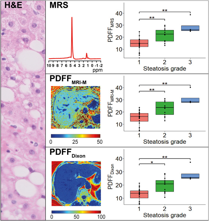
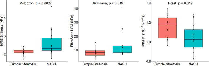
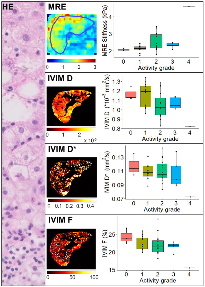
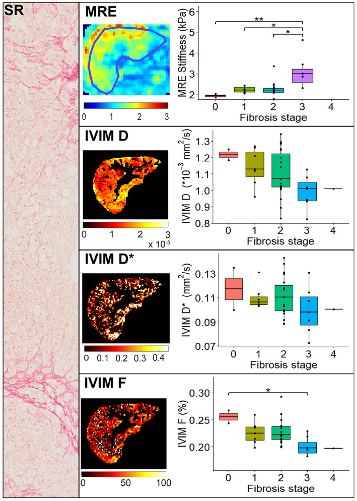
Histological steatosis grades were compared to proton density fat fraction measured by MRS (PDFF ), magnitude-based MRI (PDFF ), and three-point Dixon (PDFF ), as well as FibroScan® controlled attenuation parameter (CAP). Fibrosis and disease activity were compared to IVIM and MRE. FibroScan® liver stiffness measurements were compared to fibrosis levels. Diagnostic performance of all imaging parameters was determined for distinction between simple steatosis and nonalcoholic steatohepatitis (NASH).
Spearman's rank test, Kruskal-Wallis test, Dunn's post-hoc test with Holm-Bonferroni P-value adjustment, receiver operating characteristic curve analysis. A P-value <0.05 was considered statistically significant.
Histological steatosis grade correlated significantly with PDFF (r = 0.66, P < 0.001), PDFF (r = 0.68, P < 0.001), and PDFF (r = 0.67, P < 0.001), whereas no correlation was found with CAP. MRE and IVIM diffusion and perfusion significantly correlated with disease activity (r = 0.55, P < 0.001, r = -0.40, P = 0.016, r = -0.37, P = 0.027, respectively) and fibrosis (r = 0.55, P < 0.001, r = -0.46, P = 0.0051; r = -0.53, P < 0.001, respectively). MRE and IVIM diffusion had the highest area-under-the-curve for distinction between simple steatosis and NASH (0.79 and 0.73, respectively).
Multiparametric MRI is a promising method for noninvasive, accurate, and sensitive distinction between simple hepatic steatosis and NASH, as well as for the assessment of steatosis and fibrosis severity.
2 TECHNICAL EFFICACY: 2.
非酒精性脂肪性肝病(NAFLD)的疾病分层和监测迫切需要非侵入性诊断方法。多参数磁共振成像(MRI)是一种评估肝脂肪变性、炎症和纤维化的有前途的技术,有可能实现对具有活跃和晚期 NAFLD 的个体的非侵入性识别。
通过与组织学评分相比,检查多参数 MRI 评估 NAFLD 疾病谱中疾病严重程度的诊断性能。
前瞻性、队列研究。
37 例 NAFLD 患者。
磁场强度/序列:3.0T 多参数 MRI 包括磁共振波谱(MRS)多回波激发回波采集模式、基于幅度的和三点 Dixon 使用二维多回波梯度回波、使用广义多shot 梯度回波序列的磁共振弹性成像(MRE)和使用多切片扩散加权单次激发回波平面序列的体素内不相干运动(IVIM)。
组织学脂肪变性等级与质子密度脂肪分数(MRS 测量的 PDFF)、基于幅度的 MRI(PDFF)和三点 Dixon(PDFF)以及 FibroScan®受控衰减参数(CAP)进行比较。纤维化和疾病活动与 IVIM 和 MRE 进行比较。FibroScan®肝硬度测量与纤维化水平进行比较。确定所有成像参数的诊断性能,以区分单纯性脂肪变性和非酒精性脂肪性肝炎(NASH)。
Spearman 秩检验、Kruskal-Wallis 检验、Dunn 事后检验与 Holm-Bonferroni P 值调整、受试者工作特征曲线分析。P 值<0.05 被认为具有统计学意义。
组织学脂肪变性等级与 PDFF(r = 0.66,P<0.001)、PDFF(r = 0.68,P<0.001)和 PDFF(r = 0.67,P<0.001)显著相关,而与 CAP 无相关性。MRE 和 IVIM 扩散和灌注与疾病活动(r = 0.55,P<0.001,r = -0.40,P = 0.016,r = -0.37,P = 0.027)和纤维化(r = 0.55,P<0.001,r = -0.46,P = 0.0051;r = -0.53,P<0.001)显著相关。MRE 和 IVIM 扩散在区分单纯性脂肪变性和 NASH 方面具有最高的曲线下面积(0.79 和 0.73)。
多参数 MRI 是一种有前途的非侵入性、准确和敏感的方法,可用于区分单纯性肝脂肪变性和 NASH,以及评估脂肪变性和纤维化的严重程度。
2 技术功效:2.