• 文献检索
  • 文档翻译
  • 深度研究
  • 学术资讯
  • Suppr Zotero 插件Zotero 插件
  • 邀请有礼
  • 套餐&价格
  • 历史记录
应用&插件
Suppr Zotero 插件Zotero 插件浏览器插件Mac 客户端Windows 客户端微信小程序
定价
高级版会员购买积分包购买API积分包
服务
文献检索文档翻译深度研究API 文档MCP 服务
关于我们
关于 Suppr公司介绍联系我们用户协议隐私条款
关注我们

Suppr 超能文献

核心技术专利:CN118964589B侵权必究
粤ICP备2023148730 号-1Suppr @ 2026

文献检索

告别复杂PubMed语法,用中文像聊天一样搜索,搜遍4000万医学文献。AI智能推荐,让科研检索更轻松。

立即免费搜索

文件翻译

保留排版,准确专业,支持PDF/Word/PPT等文件格式,支持 12+语言互译。

免费翻译文档

深度研究

AI帮你快速写综述,25分钟生成高质量综述,智能提取关键信息,辅助科研写作。

立即免费体验

晚期下颌骨放射性骨坏死患者的放射性软组织损伤:放射性骨坏死病变周围各种软组织问题的初步评估与处理

Radiation-Induced Soft Tissue Injuries in Patients With Advanced Mandibular Osteoradionecrosis: A Preliminary Evaluation and Management of Various Soft Tissue Problems Around Radiation-Induced Osteonecrosis Lesions.

作者信息

Ma Chunyue, Gao Weijin, Liu Zhonglong, Zhu Dan, Zhu Fengshuo, Li Xiaoguang, He Yue

机构信息

Department of Oral Maxillofacial-Head and Neck Oncology, Shanghai Ninth People's Hospital, School of Medicine, Shanghai Jiao Tong University, Shanghai, China.

Department of Oral and Maxillofacial Surgery, First Affiliated Hospital of Wenzhou Medical University, Wenzhou, China.

出版信息

Front Oncol. 2021 Apr 28;11:641061. doi: 10.3389/fonc.2021.641061. eCollection 2021.

DOI:10.3389/fonc.2021.641061
PMID:33996554
原文链接:https://pmc.ncbi.nlm.nih.gov/articles/PMC8113699/
Abstract

OBJECTIVES

Radiation-induced soft-tissue injuries (STIs) in mandibular osteoradionecrosis (ORN) are not well studied regarding their correlations with nearby bone lesions. The aim of this study is to investigate the severity of radiation-induced STIs in advanced mandibular ORN and its relationship with hard-tissue damage and postoperative outcomes.

METHODS

A retrospective study was performed in our institution from January 2017 to December 2019. Aside from demographic factors, the associations between the triad ORN variables (irradiation doses, ORN stages, ORN sizes) and radiation-related STI factors, vascular characteristics, and postoperative functional recovery were assessed. In addition, the severity of STI was also compared with treatment outcomes. Such correlations were established both univariate and multivariable analyses.

RESULTS

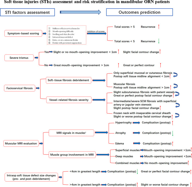
A total number of 47 patients were included. The median follow-up reached 27 months. Nasopharyngeal cancer was the histology type among most patients (n = 21, 44.7%). The median irradiation doses reached 62 Gy (range, 40-110 Gy). For STI, the symptom scoring equaled an average of 5.4 (range from 1 to 12), indicative of the severity of STI problems. During preoperative MRI examinations, signs of hypertrophy or edema (n = 41, 87.2%) were frequently discerned. Most patients (n = 23, 48.9%) also had extensive muscular fibrosis and infection, which required further debridement and scar release. Surprisingly, most STI factors, except cervical fibrosis (p = 0.02), were not in parallel with the ORN levels. Even the intraoperative soft-tissue defect changes could not be extrapolated by the extent of ORN damage (p = 0.096). Regarding the outcomes, a low recurrence rate (n = 3, 6.9%) was reported. In terms of soft tissue-related factors, we found a strong correlation (p = 0.004) between symptom scores and recurrence. In addition, when taking trismus into consideration, both improvements in mouth-opening distance (p < 0.001) and facial contour changes (p = 0.004) were adversely affected. Correlations were also observed between the intraoperative soft-tissue defect changes and complications (p = 0.024), indicative of the importance of STI evaluation and management.

CONCLUSIONS

The coexistence of hard- and soft-tissue damage in radiation-induced advanced mandibular ORN patients reminds surgeons of the significance in assessing both aspects. It is necessary to take the same active measures to evaluate and repair both severe STIs and ORN bone lesions.

摘要

目的

关于下颌骨放射性骨坏死(ORN)中辐射诱导的软组织损伤(STIs)与附近骨病变的相关性,目前尚未得到充分研究。本研究旨在调查晚期下颌骨ORN中辐射诱导的STIs的严重程度及其与硬组织损伤和术后结果的关系。

方法

2017年1月至2019年12月在本机构进行了一项回顾性研究。除人口统计学因素外,评估了ORN三联征变量(照射剂量、ORN分期、ORN大小)与辐射相关的STI因素、血管特征和术后功能恢复之间的关联。此外,还将STI的严重程度与治疗结果进行了比较。通过单变量和多变量分析建立了此类相关性。

结果

共纳入47例患者。中位随访时间达27个月。大多数患者(n = 21,44.7%)的组织学类型为鼻咽癌。中位照射剂量达62 Gy(范围40 - 110 Gy)。对于STI,症状评分平均为5.4(范围1至12),表明STI问题的严重程度。术前MRI检查时,肥大或水肿迹象(n = 41,87.2%)经常被发现。大多数患者(n = 23,48.9%)还存在广泛的肌肉纤维化和感染,需要进一步清创和松解瘢痕。令人惊讶的是,除颈部纤维化外(p = 0.02),大多数STI因素与ORN水平并不平行。即使术中软组织缺损变化也不能通过ORN损伤程度推断(p = 0.096)。关于结果,报告的复发率较低(n = 3,6.9%)。在软组织相关因素方面,我们发现症状评分与复发之间存在强相关性(p = 0.004)。此外,考虑到牙关紧闭时,开口距离的改善(p < 0.001)和面部轮廓变化(p = 0.004)均受到不利影响。术中软组织缺损变化与并发症之间也存在相关性(p = 0.024),表明STI评估和管理的重要性。

结论

辐射诱导的晚期下颌骨ORN患者硬组织和软组织损伤并存,提醒外科医生评估这两个方面的重要性。对严重的STIs和ORN骨病变采取同样积极的评估和修复措施是必要的。

https://cdn.ncbi.nlm.nih.gov/pmc/blobs/88e7/8113699/fdea0474782f/fonc-11-641061-g006.jpg
https://cdn.ncbi.nlm.nih.gov/pmc/blobs/88e7/8113699/f8f74d16a181/fonc-11-641061-g001.jpg
https://cdn.ncbi.nlm.nih.gov/pmc/blobs/88e7/8113699/74d29b480b12/fonc-11-641061-g002.jpg
https://cdn.ncbi.nlm.nih.gov/pmc/blobs/88e7/8113699/85e5aea03c0a/fonc-11-641061-g003.jpg
https://cdn.ncbi.nlm.nih.gov/pmc/blobs/88e7/8113699/961a3e0d2fd4/fonc-11-641061-g004.jpg
https://cdn.ncbi.nlm.nih.gov/pmc/blobs/88e7/8113699/97b6a1621237/fonc-11-641061-g005.jpg
https://cdn.ncbi.nlm.nih.gov/pmc/blobs/88e7/8113699/fdea0474782f/fonc-11-641061-g006.jpg

相似文献

1
Radiation-Induced Soft Tissue Injuries in Patients With Advanced Mandibular Osteoradionecrosis: A Preliminary Evaluation and Management of Various Soft Tissue Problems Around Radiation-Induced Osteonecrosis Lesions.晚期下颌骨放射性骨坏死患者的放射性软组织损伤:放射性骨坏死病变周围各种软组织问题的初步评估与处理
Front Oncol. 2021 Apr 28;11:641061. doi: 10.3389/fonc.2021.641061. eCollection 2021.
2
Risk factors and dose-effect relationship for mandibular osteoradionecrosis in oral and oropharyngeal cancer patients.口腔和口咽癌患者下颌骨放射性骨坏死的危险因素及剂量-效应关系。
Int J Radiat Oncol Biol Phys. 2009 Nov 15;75(4):1084-91. doi: 10.1016/j.ijrobp.2008.12.052. Epub 2009 Mar 26.
3
Quantitative Dynamic Contrast-Enhanced MRI Identifies Radiation-Induced Vascular Damage in Patients With Advanced Osteoradionecrosis: Results of a Prospective Study.定量动态对比增强 MRI 识别晚期放射性骨坏死患者的血管损伤:一项前瞻性研究的结果。
Int J Radiat Oncol Biol Phys. 2020 Dec 1;108(5):1319-1328. doi: 10.1016/j.ijrobp.2020.07.029. Epub 2020 Jul 23.
4
Frequency of osteoradionecrosis of the lower jaw after radiotherapy of oral cancer patients correlated with dosimetric parameters and other risk factors.口腔癌患者接受放射治疗后下颌骨放射性骨坏死的频率与剂量学参数及其他危险因素相关。
Head Face Med. 2022 Feb 26;18(1):7. doi: 10.1186/s13005-022-00311-8.
5
Radiotherapy-induced mandibular bone complications.放射治疗引起的下颌骨并发症。
Cancer Treat Rev. 2002 Feb;28(1):65-74. doi: 10.1053/ctrv.2002.0254.
6
Osteoradionecrosis of the mandible: treatment outcomes and factors influencing the progress of osteoradionecrosis.下颌骨放射性骨坏死:治疗结果及影响放射性骨坏死进展的因素
J Oral Maxillofac Surg. 2009 Jul;67(7):1378-86. doi: 10.1016/j.joms.2009.02.008.
7
Osteoradionecrosis of the mandible after radiotherapy for head and neck cancer: risk factors and dose-volume correlations.头颈部癌症放疗后下颌骨放射性骨坏死:危险因素和剂量-体积相关性。
Acta Oncol. 2019 Oct;58(10):1373-1377. doi: 10.1080/0284186X.2019.1643037. Epub 2019 Jul 31.
8
Osteoradionecrosis of the Jaw-Comparison between Bone and Soft Tissue Injury and Their Influence on Surgical Outcomes-A Retrospective Cohort Study.颌骨放射性骨坏死——骨与软组织损伤的比较及其对手术结果的影响——一项回顾性队列研究
Diagnostics (Basel). 2023 Jan 18;13(3):366. doi: 10.3390/diagnostics13030366.
9
Incidence and Risk Factors for Extremity Osteoradionecrosis after Limb-Sparing Surgery and Adjuvant Radiotherapy.保肢手术及辅助放疗后肢体放射性骨坏死的发病率及危险因素
Cancers (Basel). 2023 Apr 17;15(8):2339. doi: 10.3390/cancers15082339.
10
Surgical management of severe osteoradionecrosis of the mandibular bone by using double free flap reconstruction.采用双游离皮瓣重建术治疗下颌骨严重放射性骨坏死。
J Craniomaxillofac Surg. 2018 Jan;46(1):148-154. doi: 10.1016/j.jcms.2017.09.025. Epub 2017 Nov 2.

引用本文的文献

1
Pathological Fractures of the Mandible: Our Department's 15-Year Experience.下颌骨病理性骨折:我们科室15年的经验
Diagnostics (Basel). 2025 May 12;15(10):1216. doi: 10.3390/diagnostics15101216.
2
De-escalated radiation for human papillomavirus virus-related oropharyngeal cancer: evolving paradigms and future strategies.人乳头瘤病毒相关口咽癌的降级放疗:不断演变的模式与未来策略
Front Oncol. 2023 Jul 27;13:1175578. doi: 10.3389/fonc.2023.1175578. eCollection 2023.
3
Osteoradionecrosis of the Jaw-Comparison between Bone and Soft Tissue Injury and Their Influence on Surgical Outcomes-A Retrospective Cohort Study.

本文引用的文献

1
Head and neck squamous cell carcinoma.头颈部鳞状细胞癌
Nat Rev Dis Primers. 2020 Nov 26;6(1):92. doi: 10.1038/s41572-020-00224-3.
2
Radiation Therapy and Mandibular Osteoradionecrosis: State of the Art.放射治疗与下颌骨放射性骨坏死:现状。
Curr Oncol Rep. 2020 Jul 9;22(9):89. doi: 10.1007/s11912-020-00954-3.
3
Upper body motor function and swallowing impairments and its association in survivors of head and neck cancer: A cross-sectional study.头颈部癌症幸存者的上半身运动功能和吞咽障碍及其相关性:一项横断面研究。
颌骨放射性骨坏死——骨与软组织损伤的比较及其对手术结果的影响——一项回顾性队列研究
Diagnostics (Basel). 2023 Jan 18;13(3):366. doi: 10.3390/diagnostics13030366.
4
Modified Gap Arthroplasty for Temporomandibular Joint Ankylosis Following Radiotherapy for Rhabdomyosarcoma: Report of an Unusual Case and Brief Literature Review.横纹肌肉瘤放疗后颞下颌关节强直的改良间隙关节成形术:1例罕见病例报告及文献综述
Front Oncol. 2021 Nov 26;11:784690. doi: 10.3389/fonc.2021.784690. eCollection 2021.
PLoS One. 2020 Jun 19;15(6):e0234467. doi: 10.1371/journal.pone.0234467. eCollection 2020.
4
Radiation-Induced Skin Fibrosis: Pathogenesis, Current Treatment Options, and Emerging Therapeutics.辐射诱导的皮肤纤维化:发病机制、当前治疗选择及新兴疗法
Ann Plast Surg. 2019 Oct;83(4S Suppl 1):S59-S64. doi: 10.1097/SAP.0000000000002098.
5
Chinese expert group consensus on diagnosis and clinical management of osteoradionecrosis of the mandible.中国专家关于下颌骨放射性骨坏死的诊断和临床管理的共识。
Int J Oral Maxillofac Surg. 2020 Mar;49(3):411-419. doi: 10.1016/j.ijom.2019.06.015. Epub 2019 Jul 26.
6
Osteoradionecrosis, an increasing indication for microvascular head and neck reconstruction.放射性骨坏死,一种越来越常见的需要进行头颈部微血管重建的病症。
Int J Oral Maxillofac Surg. 2020 Jan;49(1):1-6. doi: 10.1016/j.ijom.2019.06.009. Epub 2019 Jul 8.
7
The value of reconstructive surgery in the management of refractory jaw osteoradionecrosis: a single-center 10-year experience.重建手术在难治性颌骨放射性骨坏死治疗中的价值:单中心 10 年经验。
Int J Oral Maxillofac Surg. 2019 Nov;48(11):1398-1404. doi: 10.1016/j.ijom.2019.06.007. Epub 2019 Jun 18.
8
Radiation dose to the masseter and medial pterygoid muscle in relation to trismus after chemoradiotherapy for advanced head and neck cancer.头颈部癌症放化疗后咀嚼肌和翼内肌张口困难与辐射剂量的关系。
Head Neck. 2019 May;41(5):1387-1394. doi: 10.1002/hed.25573. Epub 2019 Jan 16.
9
Documentation and incidence of late effects and screening recommendations for adolescent and young adult head and neck cancer survivors treated with radiotherapy.青少年和年轻成年头颈部癌症幸存者放疗后晚期效应的记录和发生率,以及筛查建议。
Support Care Cancer. 2019 Jul;27(7):2609-2616. doi: 10.1007/s00520-018-4559-5. Epub 2018 Nov 22.
10
Radiographic characteristics of chronic diffuse sclerosing osteomyelitis/tendoperiostitis of the mandible: A comparison with chronic suppurative osteomyelitis and osteoradionecrosis.下颌骨慢性弥漫性硬化性骨髓炎/骨膜骨膜炎的影像学特征:与慢性化脓性骨髓炎和放射性骨坏死的比较。
J Craniomaxillofac Surg. 2018 Sep;46(9):1631-1636. doi: 10.1016/j.jcms.2018.06.015. Epub 2018 Jun 30.