• 文献检索
  • 文档翻译
  • 深度研究
  • 学术资讯
  • Suppr Zotero 插件Zotero 插件
  • 邀请有礼
  • 套餐&价格
  • 历史记录
应用&插件
Suppr Zotero 插件Zotero 插件浏览器插件Mac 客户端Windows 客户端微信小程序
定价
高级版会员购买积分包购买API积分包
服务
文献检索文档翻译深度研究API 文档MCP 服务
关于我们
关于 Suppr公司介绍联系我们用户协议隐私条款
关注我们

Suppr 超能文献

核心技术专利:CN118964589B侵权必究
粤ICP备2023148730 号-1Suppr @ 2026

文献检索

告别复杂PubMed语法,用中文像聊天一样搜索,搜遍4000万医学文献。AI智能推荐,让科研检索更轻松。

立即免费搜索

文件翻译

保留排版,准确专业,支持PDF/Word/PPT等文件格式,支持 12+语言互译。

免费翻译文档

深度研究

AI帮你快速写综述,25分钟生成高质量综述,智能提取关键信息,辅助科研写作。

立即免费体验

深度学习通过分析上下文特征实现组织学图像中异体移植细胞的个体分类。

Deep Learning Enables Individual Xenograft Cell Classification in Histological Images by Analysis of Contextual Features.

机构信息

Biomedical Imaging Group, School of Engineering, Ecole Polytechnique Fédéralé de Lausanne (EPFL), Lausanne, Switzerland.

EPFL Center for Imaging, Ecole Polytechnique Fédéralé de Lausanne (EPFL), Lausanne, Switzerland.

出版信息

J Mammary Gland Biol Neoplasia. 2021 Jun;26(2):101-112. doi: 10.1007/s10911-021-09485-4. Epub 2021 May 17.

DOI:10.1007/s10911-021-09485-4
PMID:33999331
原文链接:https://pmc.ncbi.nlm.nih.gov/articles/PMC8236058/
Abstract

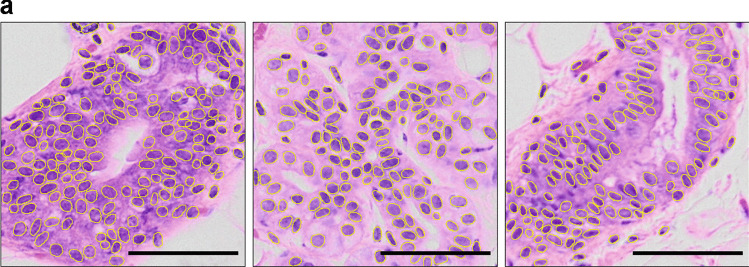
Patient-Derived Xenografts (PDXs) are the preclinical models which best recapitulate inter- and intra-patient complexity of human breast malignancies, and are also emerging as useful tools to study the normal breast epithelium. However, data analysis generated with such models is often confounded by the presence of host cells and can give rise to data misinterpretation. For instance, it is important to discriminate between xenografted and host cells in histological sections prior to performing immunostainings. We developed Single Cell Classifier (SCC), a data-driven deep learning-based computational tool that provides an innovative approach for automated cell species discrimination based on a multi-step process entailing nuclei segmentation and single cell classification. We show that human and murine cell contextual features, more than cell-intrinsic ones, can be exploited to discriminate between cell species in both normal and malignant tissues, yielding up to 96% classification accuracy. SCC will facilitate the interpretation of H&E- and DAPI-stained histological sections of xenografted human-in-mouse tissues and it is open to new in-house built models for further applications. SCC is released as an open-source plugin in ImageJ/Fiji available at the following link: https://github.com/Biomedical-Imaging-Group/SingleCellClassifier .

摘要

患者来源异种移植(PDX)是最能重现人类乳腺癌患者间和患者内复杂性的临床前模型,也正在成为研究正常乳腺上皮的有用工具。然而,使用此类模型生成的数据分析常常受到宿主细胞的干扰,并可能导致数据解读错误。例如,在进行免疫染色之前,有必要在组织切片中区分异种移植细胞和宿主细胞。我们开发了单细胞分类器(SCC),这是一种基于深度学习的数据驱动计算工具,为基于涉及核分割和单细胞分类的多步骤过程的自动细胞物种区分提供了一种创新方法。我们表明,人类和鼠类细胞的上下文特征,而不是细胞内在特征,可以用于区分正常和恶性组织中的细胞物种,分类准确率高达 96%。SCC 将有助于解释异种移植人源化小鼠组织的 H&E 和 DAPI 染色组织学切片,并可通过新的内部构建模型进行进一步应用。SCC 作为 ImageJ/Fiji 的开源插件发布,可在以下链接获得:https://github.com/Biomedical-Imaging-Group/SingleCellClassifier。

https://cdn.ncbi.nlm.nih.gov/pmc/blobs/ba50/8236058/ecaeb041e6fb/10911_2021_9485_Fig6_HTML.jpg
https://cdn.ncbi.nlm.nih.gov/pmc/blobs/ba50/8236058/3501795968c9/10911_2021_9485_Fig1_HTML.jpg
https://cdn.ncbi.nlm.nih.gov/pmc/blobs/ba50/8236058/e1fc10e7d225/10911_2021_9485_Fig2_HTML.jpg
https://cdn.ncbi.nlm.nih.gov/pmc/blobs/ba50/8236058/f7f080c4d0f4/10911_2021_9485_Fig3_HTML.jpg
https://cdn.ncbi.nlm.nih.gov/pmc/blobs/ba50/8236058/d686210d9023/10911_2021_9485_Fig4_HTML.jpg
https://cdn.ncbi.nlm.nih.gov/pmc/blobs/ba50/8236058/fc328a9c8e19/10911_2021_9485_Fig5_HTML.jpg
https://cdn.ncbi.nlm.nih.gov/pmc/blobs/ba50/8236058/ecaeb041e6fb/10911_2021_9485_Fig6_HTML.jpg

相似文献

1
Deep Learning Enables Individual Xenograft Cell Classification in Histological Images by Analysis of Contextual Features.深度学习通过分析上下文特征实现组织学图像中异体移植细胞的个体分类。
J Mammary Gland Biol Neoplasia. 2021 Jun;26(2):101-112. doi: 10.1007/s10911-021-09485-4. Epub 2021 May 17.
2
A Pan-Cancer Patient-Derived Xenograft Histology Image Repository with Genomic and Pathologic Annotations Enables Deep Learning Analysis.Pan-Cancer 患者来源异种移植组织病理学图像库与基因组和病理学注释相结合,可实现深度学习分析。
Cancer Res. 2024 Jul 2;84(13):2060-2072. doi: 10.1158/0008-5472.CAN-23-1349.
3
MaskMitosis: a deep learning framework for fully supervised, weakly supervised, and unsupervised mitosis detection in histopathology images.MaskMitosis:一种深度学习框架,用于在组织病理学图像中进行全监督、弱监督和无监督的有丝分裂检测。
Med Biol Eng Comput. 2020 Jul;58(7):1603-1623. doi: 10.1007/s11517-020-02175-z. Epub 2020 May 22.
4
Mammographic image classification with deep fusion learning.基于深度融合学习的乳腺 X 线图像分类。
Sci Rep. 2020 Sep 1;10(1):14361. doi: 10.1038/s41598-020-71431-x.
5
Normalization of HE-stained histological images using cycle consistent generative adversarial networks.使用循环一致生成对抗网络对 HE 染色组织学图像进行归一化。
Diagn Pathol. 2021 Aug 6;16(1):71. doi: 10.1186/s13000-021-01126-y.
6
Breast cancer pathological image classification based on deep learning.基于深度学习的乳腺癌病理图像分类。
J Xray Sci Technol. 2020;28(4):727-738. doi: 10.3233/XST-200658.
7
Robust detection and segmentation of cell nuclei in biomedical images based on a computational topology framework.基于计算拓扑学框架的生物医学图像中细胞核的稳健检测和分割。
Med Image Anal. 2017 May;38:90-103. doi: 10.1016/j.media.2017.02.009. Epub 2017 Mar 6.
8
A deep learning-based automated image analysis for histological evaluation of broiler pectoral muscle.基于深度学习的肉鸡胸肌组织学评估自动化图像分析。
Poult Sci. 2023 Aug;102(8):102792. doi: 10.1016/j.psj.2023.102792. Epub 2023 May 19.
9
A knowledge-driven feature learning and integration method for breast cancer diagnosis on multi-sequence MRI.基于知识的多序列 MRI 乳腺癌诊断的特征学习与融合方法
Magn Reson Imaging. 2020 Jun;69:40-48. doi: 10.1016/j.mri.2020.03.001. Epub 2020 Mar 12.
10
NucleiSegNet: Robust deep learning architecture for the nuclei segmentation of liver cancer histopathology images.细胞核分割网络:用于肝癌组织病理学图像细胞核分割的强大深度学习架构。
Comput Biol Med. 2021 Jan;128:104075. doi: 10.1016/j.compbiomed.2020.104075. Epub 2020 Nov 3.

引用本文的文献

1
Evolution and Self-renewal of the Journal of Mammary Gland Biology and Neoplasia.《乳腺生物学与肿瘤学杂志》的发展与自我更新
J Mammary Gland Biol Neoplasia. 2021 Sep;26(3):217-220. doi: 10.1007/s10911-021-09500-8. Epub 2021 Oct 14.

本文引用的文献

1
IL6/STAT3 Signaling Hijacks Estrogen Receptor α Enhancers to Drive Breast Cancer Metastasis.IL6/STAT3 信号通路劫持雌激素受体 α 增强子以驱动乳腺癌转移。
Cancer Cell. 2020 Sep 14;38(3):412-423.e9. doi: 10.1016/j.ccell.2020.06.007. Epub 2020 Jul 16.
2
Characterization of circulating breast cancer cells with tumorigenic and metastatic capacity.具有致瘤和转移能力的循环乳腺癌细胞的特征。
EMBO Mol Med. 2020 Sep 7;12(9):e11908. doi: 10.15252/emmm.201911908. Epub 2020 Jul 15.
3
Tissue-resident ductal macrophages survey the mammary epithelium and facilitate tissue remodelling.
组织驻留的导管巨噬细胞对乳腺上皮进行普查,并促进组织重塑。
Nat Cell Biol. 2020 May;22(5):546-558. doi: 10.1038/s41556-020-0505-0. Epub 2020 Apr 27.
4
The secreted protease Adamts18 links hormone action to activation of the mammary stem cell niche.分泌蛋白酶 Adamts18 将激素作用与乳腺干细胞龛的激活联系起来。
Nat Commun. 2020 Mar 26;11(1):1571. doi: 10.1038/s41467-020-15357-y.
5
International evaluation of an AI system for breast cancer screening.国际乳腺癌筛查人工智能系统评估。
Nature. 2020 Jan;577(7788):89-94. doi: 10.1038/s41586-019-1799-6. Epub 2020 Jan 1.
6
Deep Learning for Whole Slide Image Analysis: An Overview.用于全切片图像分析的深度学习:综述
Front Med (Lausanne). 2019 Nov 22;6:264. doi: 10.3389/fmed.2019.00264. eCollection 2019.
7
Content-aware image restoration: pushing the limits of fluorescence microscopy.内容感知图像恢复:推动荧光显微镜的极限。
Nat Methods. 2018 Dec;15(12):1090-1097. doi: 10.1038/s41592-018-0216-7. Epub 2018 Nov 26.
8
Multi-tissue and multi-scale approach for nuclei segmentation in H&E stained images.多组织和多尺度方法用于 H&E 染色图像中的细胞核分割。
Biomed Eng Online. 2018 Jun 20;17(1):89. doi: 10.1186/s12938-018-0518-0.
9
QuPath: Open source software for digital pathology image analysis.QuPath:用于数字病理学图像分析的开源软件。
Sci Rep. 2017 Dec 4;7(1):16878. doi: 10.1038/s41598-017-17204-5.
10
Patient-derived xenograft (PDX) models in basic and translational breast cancer research.基础及转化性乳腺癌研究中的患者来源异种移植(PDX)模型
Cancer Metastasis Rev. 2016 Dec;35(4):547-573. doi: 10.1007/s10555-016-9653-x.