Inoue Hiroyuki, Ochiai Toshiya, Kubo Hidemasa, Yamamoto Yusuke, Morimura Ryo, Ikoma Hisashi, Otsuji Eigo
Division of Digestive Surgery, Department of Surgery, Kyoto Prefectual University of Medicine, Kyoto 6028566, Japan.
Department of Surgery, North Medical Center Kyoto Prefectural University of Medicine, Kyoto 6292261, Japan.
World J Clin Cases. 2021 May 16;9(14):3424-3431. doi: 10.12998/wjcc.v9.i14.3424.
Gangrenous cholecystitis is a form of acute cholecystitis which involves gangrenous alterations in the gallbladder wall and it often follows an acute and serious course. We herein report on two cases of very elderly people diagnosed early with gangrenous cholecystitis, who safely underwent laparoscopic cholecystectomy (LC) and both demonstrated a good outcome.
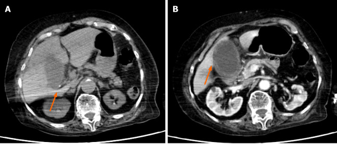
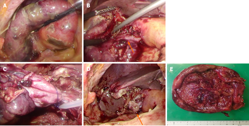
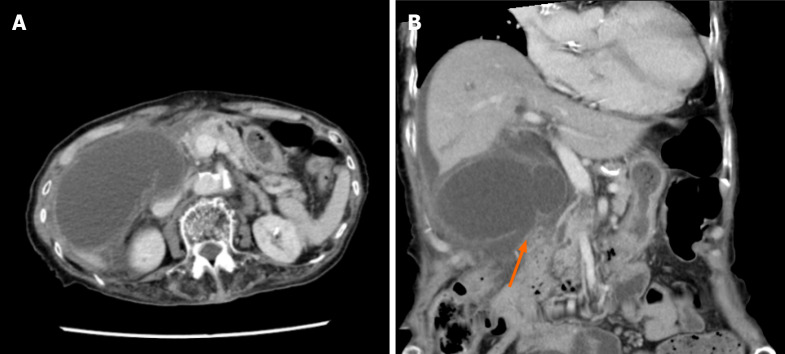
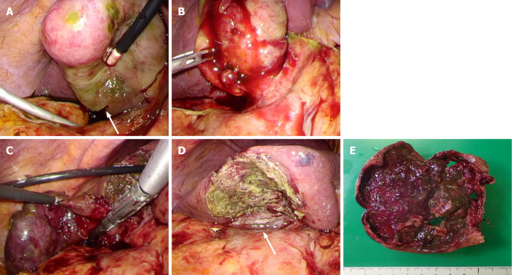
Case 1: An 89-year-old female. She underwent abdominal contrast-enhanced computed tomography (CECT) due to abdominal pain and diarrhea. Her gallbladder wall indicated the absence of contrast enhancement, thus leading to diagnosis of gangrenous cholecystitis and she therefore underwent LC. Although her gallbladder demonstrated diffuse necrosis and it was also partly perforated, she was able to be discharged without any serious complications. Case 2: A 91-year-old female. She made an emergency visit with a chief complaint of abdominal pain. Abdominal CECT revealed swelling of the gallbladder and an ambiguous continuity of the gallbladder wall. She was diagnosed with gangrenous cholecystitis and underwent LC. Her gallbladder had swelling and diffuse necrosis. Although her preoperative blood culture was positive, she showed a good outcome following surgery.
Although a definite diagnosis of gangrenous cholecystitis is difficult to make prior to surgery, if an early diagnosis can be made and appropriate treatment can be carried out, then even very elderly individuals may be discharged without major complications.
坏疽性胆囊炎是急性胆囊炎的一种形式,涉及胆囊壁的坏疽性改变,通常病程急性且严重。我们在此报告两例早期诊断为坏疽性胆囊炎的高龄患者,他们安全地接受了腹腔镜胆囊切除术(LC),且均显示出良好的预后。
病例1:一名89岁女性。因腹痛和腹泻接受腹部增强计算机断层扫描(CECT)。其胆囊壁显示无造影剂增强,从而诊断为坏疽性胆囊炎,因此接受了LC。尽管她的胆囊显示弥漫性坏死且部分穿孔,但她能够出院,没有任何严重并发症。病例2:一名91岁女性。因腹痛为主诉前来急诊。腹部CECT显示胆囊肿大,胆囊壁连续性不明确。她被诊断为坏疽性胆囊炎并接受了LC。她的胆囊肿大且弥漫性坏死。尽管她术前血培养呈阳性,但术后预后良好。
尽管术前很难对坏疽性胆囊炎做出明确诊断,但如果能早期诊断并进行适当治疗,那么即使是高龄患者也可能无重大并发症而出院。