Mental Health and Clinical Neurosciences Academic Unit, School of Medicine, University of Nottingham, Queen's Medical Centre, Nottingham NG7 2UH, United Kingdom.
Mental Health and Clinical Neurosciences Academic Unit, School of Medicine, University of Nottingham, Queen's Medical Centre, Nottingham NG7 2UH, United Kingdom.
Neuroimage Clin. 2021;31:102697. doi: 10.1016/j.nicl.2021.102697. Epub 2021 May 8.
The global incidence of traumatic brain injuries is rising, with at least 80% being classified as mild. These mild injuries are not visible on routine clinical imaging. The potential clinical role of a specific imaging biomarker be it diagnostic, prognostic or directing and monitoring progress of personalised treatment and rehabilitation has driven the exploration of several new neuroimaging modalities. This systematic review examined the evidence for magnetoencephalography (MEG) to provide an imaging biomarker in mild traumatic brain injury (mTBI).
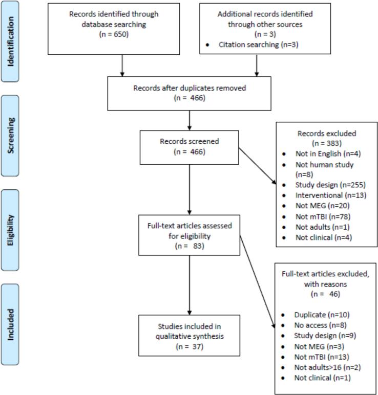
Our review was prospectively registered on PROSPERO: CRD42019151387. We searched EMBASE, MEDLINE, trial registers, PsycINFO, Cochrane Library and conference abstracts and identified 37 papers describing MEG changes in mTBI eligible for inclusion. Since meta-analysis was not possible, based on the heterogeneity of reported outcomes, we provide a narrative synthesis of results.
The two most promising MEG biomarkers are excess resting state low frequency power, and widespread connectivity changes in all frequency bands. These may represent biomarkers with potential for diagnostic application, which reflect time sensitive changes, or may be capable of offering clinically relevant prognostic information. In addition, the rich data that MEG produces are well-suited to new methods of machine learning analysis, which is now being actively explored.
MEG reveals several promising biomarkers, in the absence of structural abnormalities demonstrable with either computerised tomography or magnetic resonance imaging. This review has not identified sufficient evidence to support routine clinical use of MEG in mTBI currently. However, verifying MEG's potential would help meet an urgent clinical need within civilian, sports and military medicine.
全球创伤性脑损伤的发病率正在上升,其中至少 80%被归类为轻度损伤。这些轻度损伤在常规临床成像中不可见。特定成像生物标志物的潜在临床作用——无论是诊断、预后还是指导和监测个性化治疗和康复的进展——推动了几种新的神经影像学模式的探索。本系统评价检查了脑磁图(MEG)在轻度创伤性脑损伤(mTBI)中提供成像生物标志物的证据。
我们的综述前瞻性地在 PROSPERO 上注册:CRD42019151387。我们搜索了 EMBASE、MEDLINE、试验登记处、PsycINFO、Cochrane 图书馆和会议摘要,并确定了 37 篇描述 mTBI 中 MEG 变化的论文符合纳入标准。由于报告的结果存在异质性,不适合进行荟萃分析,因此我们提供了结果的叙述性综合。
两种最有前途的 MEG 生物标志物是静息状态下低频功率增加和所有频段的广泛连接变化。这些可能代表具有诊断应用潜力的生物标志物,反映了时间敏感的变化,或者能够提供有临床意义的预后信息。此外,MEG 产生的丰富数据非常适合机器学习分析的新方法,目前正在积极探索。
MEG 揭示了几种有前途的生物标志物,而计算机断层扫描或磁共振成像无法显示这些生物标志物。目前,本综述没有发现足够的证据支持在 mTBI 中常规使用 MEG。然而,验证 MEG 的潜力将有助于满足民用、运动和军事医学领域的迫切临床需求。