Department of Psychiatry, Brigham and Women's Hospital, Harvard Medical School, Boston, MA, USA.
Melbourne Neuropsychiatry Centre, Department of Psychiatry, The University of Melbourne and Melbourne Health, Carlton South, VIC, Australia.
Mol Psychiatry. 2021 Nov;26(11):6833-6844. doi: 10.1038/s41380-021-01128-8. Epub 2021 May 24.
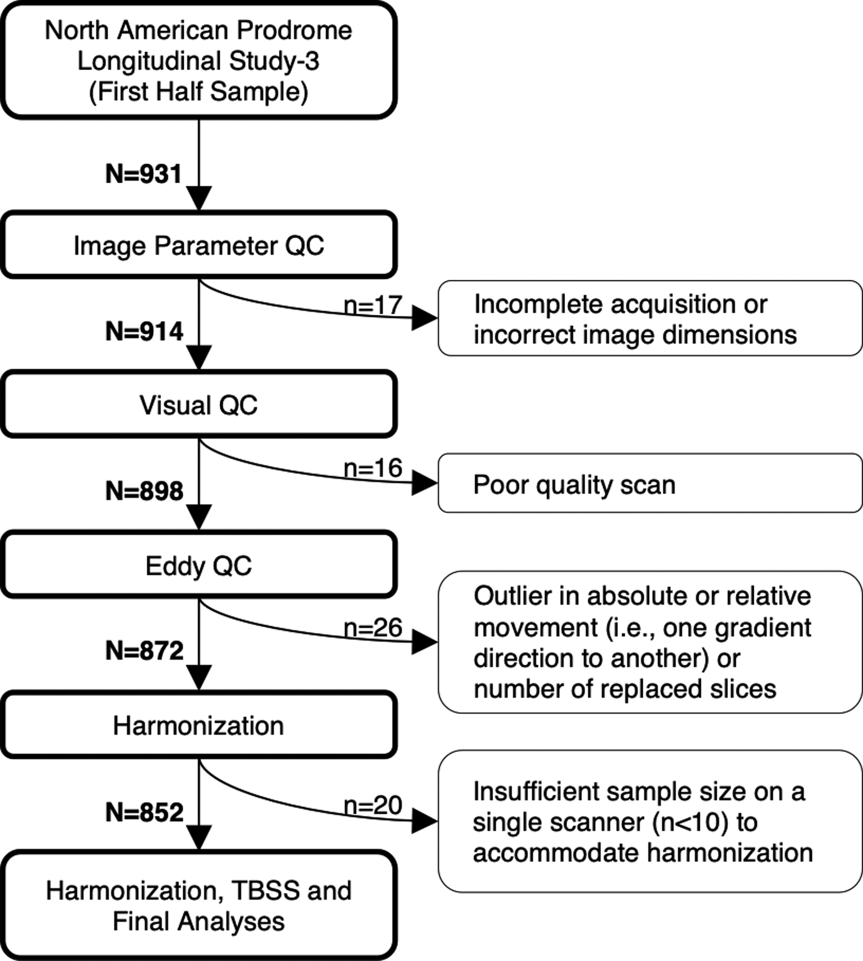
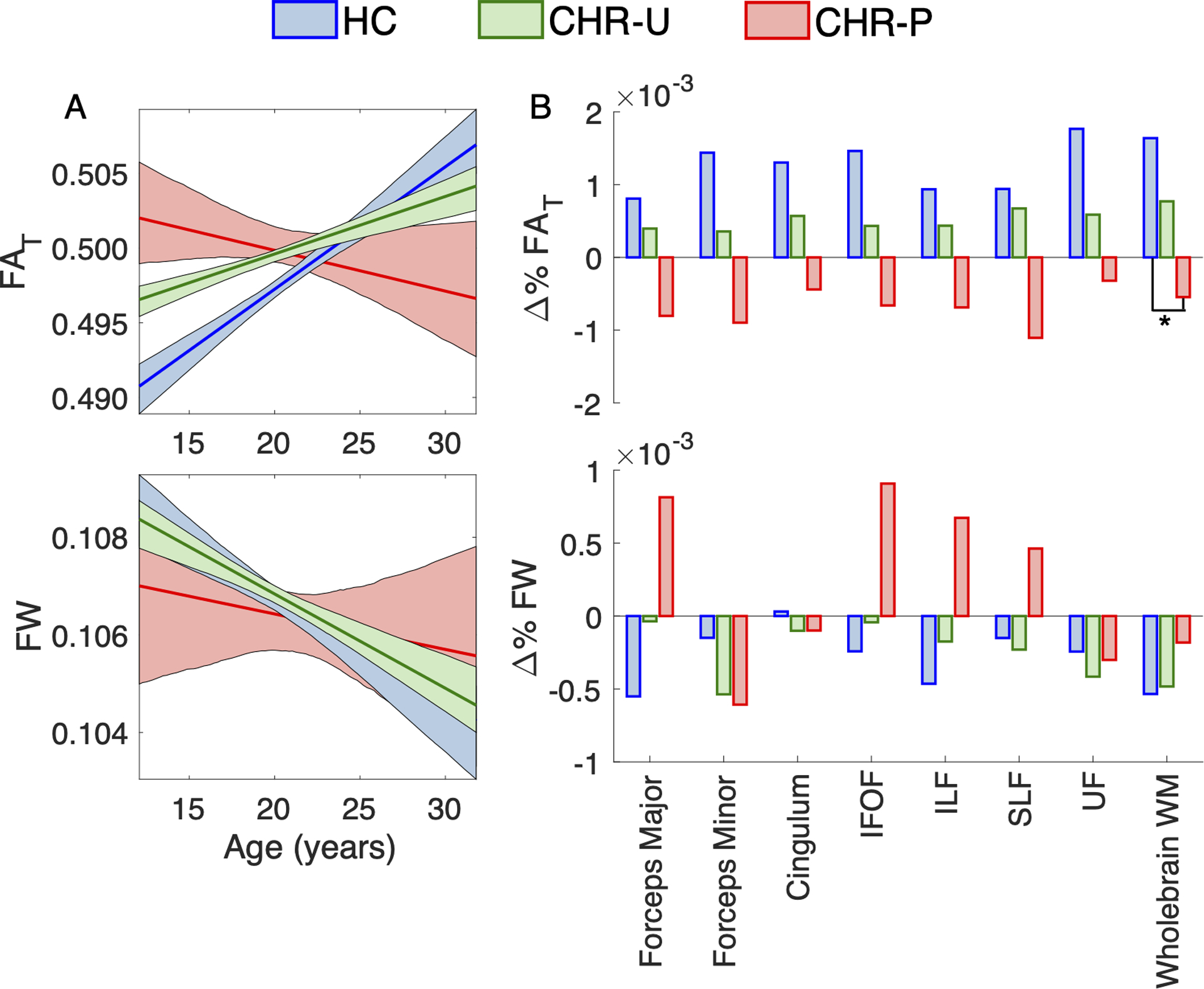
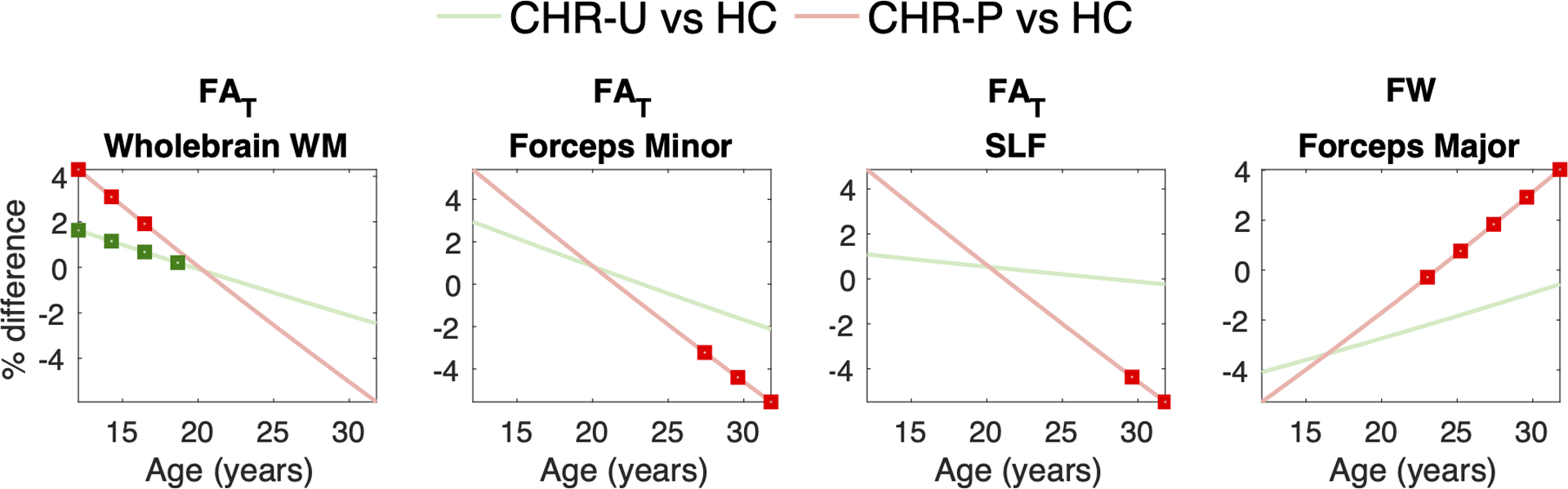
Subtle alterations in white matter microstructure are observed in youth at clinical high risk (CHR) for psychosis. However, the timing of these changes and their relationships to the emergence of psychosis remain unclear. Here, we track the evolution of white matter abnormalities in a large, longitudinal cohort of CHR individuals comprising the North American Prodrome Longitudinal Study (NAPLS-3). Multi-shell diffusion magnetic resonance imaging data were collected across multiple timepoints (1-5 over 1 year) in 286 subjects (aged 12-32 years): 25 CHR individuals who transitioned to psychosis (CHR-P; 61 scans), 205 CHR subjects with unknown transition outcome after the 1-year follow-up period (CHR-U; 596 scans), and 56 healthy controls (195 scans). Linear mixed effects models were fitted to infer the impact of age and illness-onset on variation in the fractional anisotropy of cellular tissue (FA) and the volume fraction of extracellular free water (FW). Baseline measures of white matter microstructure did not differentiate between HC, CHR-U and CHR-P individuals. However, age trajectories differed between the three groups in line with a developmental effect: CHR-P and CHR-U groups displayed higher FA in adolescence, and 4% lower FA by 30 years of age compared to controls. Furthermore, older CHR-P subjects (20+ years) displayed 4% higher FW in the forceps major (p < 0.05). Prospective analysis in CHR-P did not reveal a significant impact of illness onset on regional FA or FW, suggesting that transition to psychosis is not marked by dramatic change in white matter microstructure. Instead, clinical high risk for psychosis-regardless of transition outcome-is characterized by subtle age-related white matter changes that occur in tandem with development.
在处于精神病临床高风险(CHR)的年轻人中,观察到白质微观结构的细微改变。然而,这些变化的时间及其与精神病发作的关系尚不清楚。在这里,我们跟踪了一个大型纵向 CHR 个体队列(包括北美前驱纵向研究(NAPLS-3))中白质异常的演变。在 286 名受试者(年龄在 12-32 岁之间)中,在多个时间点(1-5 个点,为期 1 年)收集了多壳扩散磁共振成像数据:25 名过渡到精神病的 CHR 个体(CHR-P;61 次扫描),205 名在 1 年随访后未知是否过渡到精神病的 CHR 个体(CHR-U;596 次扫描),以及 56 名健康对照者(195 次扫描)。线性混合效应模型用于推断年龄和发病对细胞组织各向异性分数(FA)和细胞外游离水容积分数(FW)变化的影响。基线白质微观结构测量值无法区分 HC、CHR-U 和 CHR-P 个体。然而,这三个组的年龄轨迹不同,符合发育效应:CHR-P 和 CHR-U 组在青春期的 FA 值较高,而到 30 岁时的 FA 值比对照组低 4%。此外,年龄较大的 CHR-P 受试者(20 岁以上)在主要钳夹部的 FW 值高 4%(p<0.05)。CHR-P 的前瞻性分析并未显示发病对区域 FA 或 FW 的显著影响,这表明向精神病的过渡并非以白质微观结构的剧烈变化为标志。相反,精神病的临床高风险——无论过渡结果如何——都以与发育并行的微妙的与年龄相关的白质变化为特征。