• 文献检索
  • 文档翻译
  • 深度研究
  • 学术资讯
  • Suppr Zotero 插件Zotero 插件
  • 邀请有礼
  • 套餐&价格
  • 历史记录
应用&插件
Suppr Zotero 插件Zotero 插件浏览器插件Mac 客户端Windows 客户端微信小程序
定价
高级版会员购买积分包购买API积分包
服务
文献检索文档翻译深度研究API 文档MCP 服务
关于我们
关于 Suppr公司介绍联系我们用户协议隐私条款
关注我们

Suppr 超能文献

核心技术专利:CN118964589B侵权必究
粤ICP备2023148730 号-1Suppr @ 2026

文献检索

告别复杂PubMed语法,用中文像聊天一样搜索,搜遍4000万医学文献。AI智能推荐,让科研检索更轻松。

立即免费搜索

文件翻译

保留排版,准确专业,支持PDF/Word/PPT等文件格式,支持 12+语言互译。

免费翻译文档

深度研究

AI帮你快速写综述,25分钟生成高质量综述,智能提取关键信息,辅助科研写作。

立即免费体验

整合细胞类型特异性多组学方法揭示了DM1小鼠培养模型中胶质细胞分化程序的受损情况。

Integrative Cell Type-Specific Multi-Omics Approaches Reveal Impaired Programs of Glial Cell Differentiation in Mouse Culture Models of DM1.

作者信息

González-Barriga Anchel, Lallemant Louison, Dincã Diana M, Braz Sandra O, Polvèche Hélène, Magneron Paul, Pionneau Cédric, Huguet-Lachon Aline, Claude Jean-Baptiste, Chhuon Cerina, Guerrera Ida Chiara, Bourgeois Cyril F, Auboeuf Didier, Gourdon Geneviève, Gomes-Pereira Mário

机构信息

Sorbonne Université, Inserm, Institut de Myologie, Centre de Recherche en Myologie, Paris, France.

Inserm UMR 1163, Institut Imagine, Université Paris Cité, Paris, France.

出版信息

Front Cell Neurosci. 2021 May 5;15:662035. doi: 10.3389/fncel.2021.662035. eCollection 2021.

DOI:10.3389/fncel.2021.662035
PMID:34025359
原文链接:https://pmc.ncbi.nlm.nih.gov/articles/PMC8136287/
Abstract

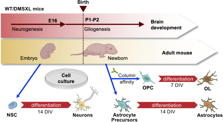
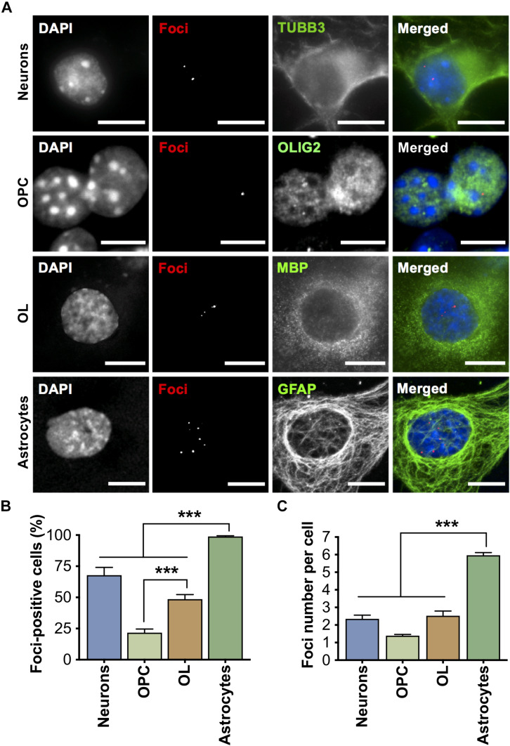
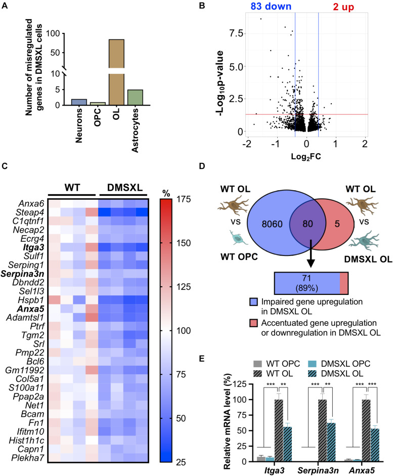
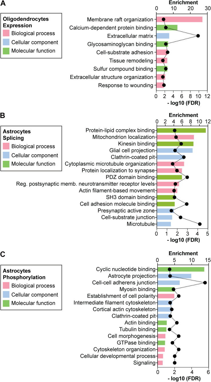
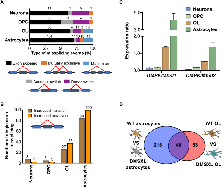
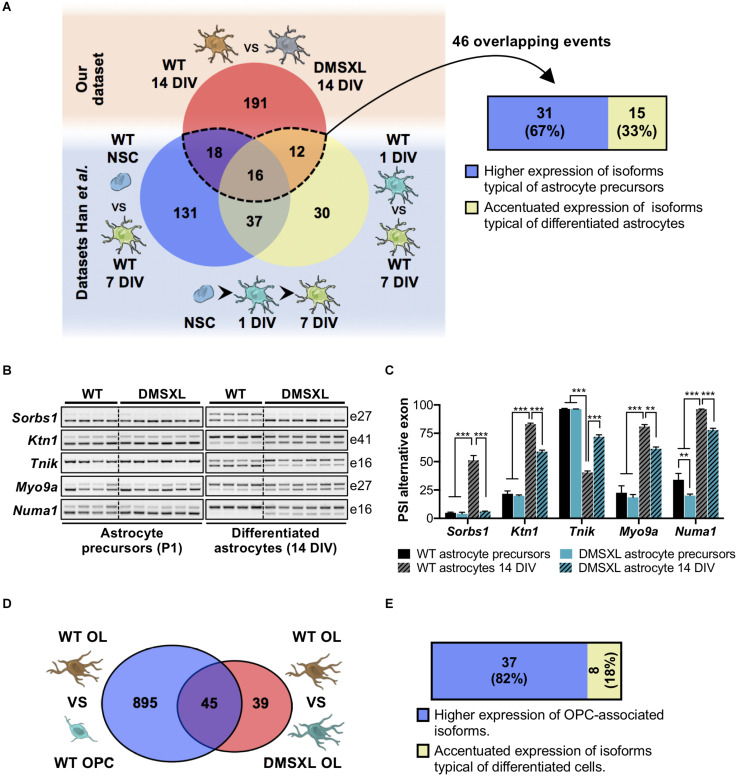
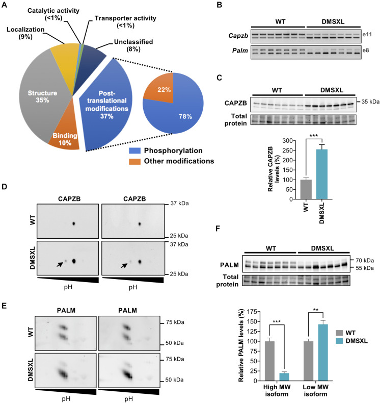
Myotonic dystrophy type 1 (DM1) is a neuromuscular disorder caused by a non-coding CTG repeat expansion in the gene. This mutation generates a toxic CUG RNA that interferes with the RNA processing of target genes in multiple tissues. Despite debilitating neurological impairment, the pathophysiological cascade of molecular and cellular events in the central nervous system (CNS) has been less extensively characterized than the molecular pathogenesis of muscle/cardiac dysfunction. Particularly, the contribution of different cell types to DM1 brain disease is not clearly understood. We first used transcriptomics to compare the impact of expanded CUG RNA on the transcriptome of primary neurons, astrocytes and oligodendrocytes derived from DMSXL mice, a transgenic model of DM1. RNA sequencing revealed more frequent expression and splicing changes in glia than neuronal cells. In particular, primary DMSXL oligodendrocytes showed the highest number of transcripts differentially expressed, while DMSXL astrocytes displayed the most severe splicing dysregulation. Interestingly, the expression and splicing defects of DMSXL glia recreated molecular signatures suggestive of impaired cell differentiation: while DMSXL oligodendrocytes failed to upregulate a subset of genes that are naturally activated during the oligodendroglia differentiation, a significant proportion of missplicing events in DMSXL oligodendrocytes and astrocytes increased the expression of RNA isoforms typical of precursor cell stages. Together these data suggest that expanded CUG RNA in glial cells affects preferentially differentiation-regulated molecular events. This hypothesis was corroborated by gene ontology (GO) analyses, which revealed an enrichment for biological processes and cellular components with critical roles during cell differentiation. Finally, we combined exon ontology with phosphoproteomics and cell imaging to explore the functional impact of CUG-associated spliceopathy on downstream protein metabolism. Changes in phosphorylation, protein isoform expression and intracellular localization in DMSXL astrocytes demonstrate the far-reaching impact of the DM1 repeat expansion on cell metabolism. Our multi-omics approaches provide insight into the mechanisms of CUG RNA toxicity in the CNS with cell type resolution, and support the priority for future research on non-neuronal mechanisms and proteomic changes in DM1 brain disease.

摘要

1型强直性肌营养不良(DM1)是一种由该基因中非编码CTG重复序列扩增引起的神经肌肉疾病。这种突变产生一种有毒的CUG RNA,它会干扰多个组织中靶基因的RNA加工过程。尽管存在使人衰弱的神经功能障碍,但与肌肉/心脏功能障碍的分子发病机制相比,中枢神经系统(CNS)中分子和细胞事件的病理生理级联反应尚未得到广泛的表征。特别是,不同细胞类型对DM1脑疾病的贡献尚不清楚。我们首先使用转录组学来比较扩增的CUG RNA对源自DMSXL小鼠(一种DM1转基因模型)的原代神经元、星形胶质细胞和少突胶质细胞转录组的影响。RNA测序显示,与神经元细胞相比,神经胶质细胞中表达和剪接变化更频繁。特别是,原代DMSXL少突胶质细胞显示出差异表达转录本数量最多,而DMSXL星形胶质细胞则表现出最严重的剪接失调。有趣的是,DMSXL神经胶质细胞的表达和剪接缺陷重现了提示细胞分化受损的分子特征:虽然DMSXL少突胶质细胞未能上调少突胶质细胞分化过程中自然激活的一部分基因,但DMSXL少突胶质细胞和星形胶质细胞中相当一部分错配剪接事件增加了前体细胞阶段典型RNA异构体的表达。这些数据共同表明,神经胶质细胞中扩增的CUG RNA优先影响分化调节的分子事件。基因本体(GO)分析证实了这一假设,该分析揭示了在细胞分化过程中起关键作用的生物过程和细胞成分的富集。最后,我们将外显子本体与磷酸蛋白质组学和细胞成像相结合,以探索CUG相关剪接病对下游蛋白质代谢的功能影响。DMSXL星形胶质细胞中磷酸化、蛋白质异构体表达和细胞内定位的变化证明了DM1重复序列扩增对细胞代谢的深远影响。我们的多组学方法以细胞类型分辨率深入了解了CUG RNA在中枢神经系统中的毒性机制,并支持未来对DM1脑疾病中非神经元机制和蛋白质组变化进行研究的优先性。

https://cdn.ncbi.nlm.nih.gov/pmc/blobs/f478/8136287/c6b372e0e4df/fncel-15-662035-g008.jpg
https://cdn.ncbi.nlm.nih.gov/pmc/blobs/f478/8136287/1d707eac7100/fncel-15-662035-g001.jpg
https://cdn.ncbi.nlm.nih.gov/pmc/blobs/f478/8136287/10a6a6efdb3c/fncel-15-662035-g002.jpg
https://cdn.ncbi.nlm.nih.gov/pmc/blobs/f478/8136287/e8decc6f5df9/fncel-15-662035-g003.jpg
https://cdn.ncbi.nlm.nih.gov/pmc/blobs/f478/8136287/d9867b3c2a4d/fncel-15-662035-g004.jpg
https://cdn.ncbi.nlm.nih.gov/pmc/blobs/f478/8136287/d9ebf918d3db/fncel-15-662035-g005.jpg
https://cdn.ncbi.nlm.nih.gov/pmc/blobs/f478/8136287/eb38bf3fd636/fncel-15-662035-g006.jpg
https://cdn.ncbi.nlm.nih.gov/pmc/blobs/f478/8136287/7dc38f4ad3b9/fncel-15-662035-g007.jpg
https://cdn.ncbi.nlm.nih.gov/pmc/blobs/f478/8136287/c6b372e0e4df/fncel-15-662035-g008.jpg

相似文献

1
Integrative Cell Type-Specific Multi-Omics Approaches Reveal Impaired Programs of Glial Cell Differentiation in Mouse Culture Models of DM1.整合细胞类型特异性多组学方法揭示了DM1小鼠培养模型中胶质细胞分化程序的受损情况。
Front Cell Neurosci. 2021 May 5;15:662035. doi: 10.3389/fncel.2021.662035. eCollection 2021.
2
Molecular, physiological, and motor performance defects in DMSXL mice carrying >1,000 CTG repeats from the human DM1 locus.携带人源 DM1 基因座 >1000 个 CTG 重复的 DMSXL 小鼠的分子、生理和运动表现缺陷。
PLoS Genet. 2012;8(11):e1003043. doi: 10.1371/journal.pgen.1003043. Epub 2012 Nov 29.
3
Transcriptome Analysis Reveals Altered Inflammatory Pathway in an Inducible Glial Cell Model of Myotonic Dystrophy Type 1.转录组分析揭示了 1 型强直性肌营养不良诱导性神经胶质细胞模型中炎症途径的改变。
Biomolecules. 2021 Jan 26;11(2):159. doi: 10.3390/biom11020159.
4
Synaptic protein dysregulation in myotonic dystrophy type 1: Disease neuropathogenesis beyond missplicing.1型强直性肌营养不良中的突触蛋白失调:超越剪接异常的疾病神经发病机制
Rare Dis. 2013 Jun 26;1:e25553. doi: 10.4161/rdis.25553. eCollection 2013.
5
Therapeutic Targeting of the GSK3β-CUGBP1 Pathway in Myotonic Dystrophy.肌强直性营养不良症中 GSK3β-CUGBP1 通路的治疗靶点。
Int J Mol Sci. 2023 Jun 26;24(13):10650. doi: 10.3390/ijms241310650.
6
A CTG repeat-selective chemical screen identifies microtubule inhibitors as selective modulators of toxic CUG RNA levels.一种 CTG 重复序列选择性化学筛选方法,可识别微管抑制剂作为毒性 CUG RNA 水平的选择性调节剂。
Proc Natl Acad Sci U S A. 2019 Oct 15;116(42):20991-21000. doi: 10.1073/pnas.1901893116. Epub 2019 Sep 30.
7
Functional and histopathological identification of the respiratory failure in a DMSXL transgenic mouse model of myotonic dystrophy.杜氏肌营养不良症(DMSXL)转基因小鼠模型呼吸衰竭的功能和组织病理学鉴定。
Dis Model Mech. 2013 May;6(3):622-31. doi: 10.1242/dmm.010512. Epub 2012 Nov 23.
8
Furamidine Rescues Myotonic Dystrophy Type I Associated Mis-Splicing through Multiple Mechanisms.呋咱甲氢龙通过多种机制挽救肌强直性营养不良 1 型相关的剪接错误。
ACS Chem Biol. 2018 Sep 21;13(9):2708-2718. doi: 10.1021/acschembio.8b00646. Epub 2018 Aug 27.
9
Chronic exercise mitigates disease mechanisms and improves muscle function in myotonic dystrophy type 1 mice.慢性运动减轻肌强直性营养不良 1 型小鼠的疾病机制并改善肌肉功能。
J Physiol. 2019 Mar;597(5):1361-1381. doi: 10.1113/JP277123. Epub 2019 Jan 30.
10
A low absolute number of expanded transcripts is involved in myotonic dystrophy type 1 manifestation in muscle.低绝对数量的扩增转录本参与了1型强直性肌营养不良症在肌肉中的表现。
Hum Mol Genet. 2016 Apr 15;25(8):1648-62. doi: 10.1093/hmg/ddw042. Epub 2016 Feb 16.

引用本文的文献

1
Muscle-specific gene editing improves molecular and phenotypic defects in a mouse model of myotonic dystrophy type 1.肌肉特异性基因编辑改善了1型强直性肌营养不良小鼠模型中的分子和表型缺陷。
Clin Transl Med. 2025 Feb;15(2):e70227. doi: 10.1002/ctm2.70227.
2
Excessive rest time during active phase is reliably detected in a mouse model of myotonic dystrophy type 1 using home cage monitoring.利用笼内监测技术,在1型强直性肌营养不良小鼠模型中能够可靠地检测到活动期的休息时间过长。
Front Behav Neurosci. 2023 Mar 2;17:1130055. doi: 10.3389/fnbeh.2023.1130055. eCollection 2023.
3
Protein Phosphorylation Alterations in Myotonic Dystrophy Type 1: A Systematic Review.

本文引用的文献

1
Transcriptome alterations in myotonic dystrophy frontal cortex.强直性肌营养不良症额叶皮质的转录组改变。
Cell Rep. 2021 Jan 19;34(3):108634. doi: 10.1016/j.celrep.2020.108634.
2
Functions of p38 MAP Kinases in the Central Nervous System.p38丝裂原活化蛋白激酶在中枢神经系统中的功能
Front Mol Neurosci. 2020 Sep 8;13:570586. doi: 10.3389/fnmol.2020.570586. eCollection 2020.
3
Microtubule organization and dynamics in oligodendrocytes, astrocytes, and microglia.少突胶质细胞、星形胶质细胞和小胶质细胞中的微管组织和动力学。
1 型肌强直性营养不良蛋白磷酸化改变:系统评价。
Int J Mol Sci. 2023 Feb 4;24(4):3091. doi: 10.3390/ijms24043091.
4
Deciphering the Complex Molecular Pathogenesis of Myotonic Dystrophy Type 1 through Omics Studies.通过组学研究破解 1 型肌强直性营养不良的复杂分子发病机制。
Int J Mol Sci. 2022 Jan 27;23(3):1441. doi: 10.3390/ijms23031441.
5
DM1 Transgenic Mice Exhibit Abnormal Neurotransmitter Homeostasis and Synaptic Plasticity in Association with RNA Foci and Mis-Splicing in the Hippocampus.DM1 转基因小鼠表现出异常的神经递质动态平衡和海马突触可塑性,与 RNA 焦点和剪接错误有关。
Int J Mol Sci. 2022 Jan 6;23(2):592. doi: 10.3390/ijms23020592.
6
Brain Pathogenesis and Potential Therapeutic Strategies in Myotonic Dystrophy Type 1.1型强直性肌营养不良症的脑发病机制及潜在治疗策略
Front Aging Neurosci. 2021 Nov 15;13:755392. doi: 10.3389/fnagi.2021.755392. eCollection 2021.
Dev Neurobiol. 2021 Apr;81(3):310-320. doi: 10.1002/dneu.22753. Epub 2020 May 9.
4
HNRNPA1-induced spliceopathy in a transgenic mouse model of myotonic dystrophy.HNRNPA1 诱导的肌强直性营养不良转基因小鼠模型中的剪接病变。
Proc Natl Acad Sci U S A. 2020 Mar 10;117(10):5472-5477. doi: 10.1073/pnas.1907297117. Epub 2020 Feb 21.
5
Consensus-based care recommendations for congenital and childhood-onset myotonic dystrophy type 1.基于共识的1型先天性和儿童期发病的强直性肌营养不良症护理建议。
Neurol Clin Pract. 2019 Oct;9(5):443-454. doi: 10.1212/CPJ.0000000000000646.
6
Real Time Videomicroscopy and Semiautomated Analysis of Brain Cell Culture Models of Trinucleotide Repeat Expansion Diseases.三核苷酸重复扩增疾病脑细胞培养模型的实时视频显微镜检查和半自动分析
Methods Mol Biol. 2020;2056:217-240. doi: 10.1007/978-1-4939-9784-8_14.
7
Deprivation of Muscleblind-Like Proteins Causes Deficits in Cortical Neuron Distribution and Morphological Changes in Dendritic Spines and Postsynaptic Densities.肌肉盲样蛋白缺失导致皮层神经元分布缺陷以及树突棘和突触后致密物的形态变化。
Front Neuroanat. 2019 Jul 30;13:75. doi: 10.3389/fnana.2019.00075. eCollection 2019.
8
Short Tandem Repeat Expansions and RNA-Mediated Pathogenesis in Myotonic Dystrophy.短串联重复扩展与肌强直性营养不良的 RNA 介导发病机制。
Int J Mol Sci. 2019 Jul 9;20(13):3365. doi: 10.3390/ijms20133365.
9
CapZ integrates several signaling pathways in response to mechanical stiffness.衔接蛋白 Z 通过整合多种信号通路对机械刚性做出响应。
J Gen Physiol. 2019 May 6;151(5):660-669. doi: 10.1085/jgp.201812199. Epub 2019 Feb 26.
10
RNA-Mediated Disease Mechanisms in Neurodegenerative Disorders.RNA 介导的神经退行性疾病发病机制。
J Mol Biol. 2019 Apr 19;431(9):1780-1791. doi: 10.1016/j.jmb.2018.12.012. Epub 2018 Dec 29.