Oh Sanghoon, Jung Wi Hoon, Kim Taekwan, Shim Geumsook, Kwon Jun Soo
Department of Psychiatry, Seoul National University College of Medicine, Seoul, South Korea.
Department of Psychiatry, Uijeongbu Eulji Medical Center, Eulji University School of Medicine, Gyeonggi-do, South Korea.
Front Psychiatry. 2021 May 7;12:659121. doi: 10.3389/fpsyt.2021.659121. eCollection 2021.
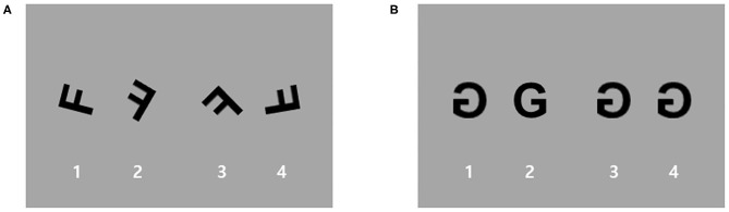
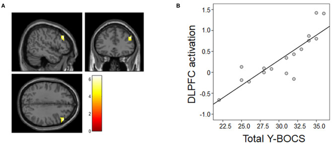
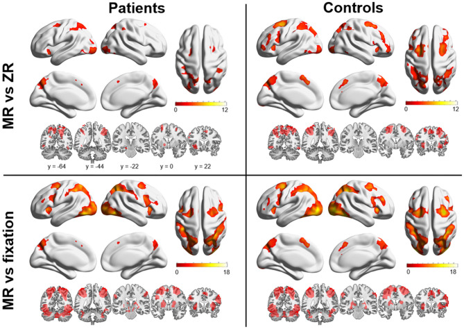
Functional neuroimaging studies have implicated alterations in frontostriatal and frontoparietal circuits in obsessive-compulsive disorder (OCD) during various tasks. To date, however, brain activation for visuospatial function in conjunction with symptoms in OCD has not been comprehensively evaluated. To elucidate the relationship between neural activity, cognitive function, and obsessive-compulsive symptoms, we investigated regional brain activation during the performance of a visuospatial task in patients with OCD using functional magnetic resonance imaging (fMRI). Seventeen medication-free patients with OCD and 21 age-, sex-, and IQ-matched healthy controls participated in this study. Functional magnetic resonance imaging data were obtained while the subjects performed a mental rotation (MR) task. Brain activation during the task was compared between the two groups using a two-sample -test. Voxel-wise whole-brain multiple regression analyses were also performed to examine the relationship between obsessive-compulsive symptom severity and neural activity during the task. The two groups did not differ in MR task performance. Both groups also showed similar task-related activation patterns in frontoparietal regions with no significant differences. Activation in the right dorsolateral prefrontal cortex in patients with OCD during the MR task was positively associated with their total Yale-Brown Obsessive-Compulsive Scale (Y-BOCS) scores. This study identified the specific brain areas associated with the interaction between symptom severity and visuospatial cognitive function during an MR task in medication-free patients with OCD. These findings may serve as potential neuromodulation targets for OCD treatment.
功能神经影像学研究表明,在各种任务中,强迫症(OCD)患者的额纹状体和额顶叶回路存在改变。然而,迄今为止,尚未全面评估强迫症患者视觉空间功能的脑激活与症状之间的关系。为了阐明神经活动、认知功能和强迫症状之间的关系,我们使用功能磁共振成像(fMRI)研究了强迫症患者在执行视觉空间任务时的脑区激活情况。17名未服用药物的强迫症患者和21名年龄、性别和智商匹配的健康对照者参与了这项研究。在受试者执行心理旋转(MR)任务时获取功能磁共振成像数据。使用双样本t检验比较两组在任务期间的脑激活情况。还进行了体素全脑多元回归分析,以检验任务期间强迫症状严重程度与神经活动之间的关系。两组在MR任务表现上没有差异。两组在额顶叶区域也表现出相似的任务相关激活模式,无显著差异。强迫症患者在MR任务期间右侧背外侧前额叶皮质的激活与其耶鲁-布朗强迫症量表(Y-BOCS)总分呈正相关。本研究确定了未服用药物的强迫症患者在MR任务期间与症状严重程度和视觉空间认知功能相互作用相关的特定脑区。这些发现可能成为强迫症治疗的潜在神经调节靶点。