Issiaka Moctar, Jamaleddine Hamza, El Belhadji Mohamed, Iro Salissou, Slimani Faiçal
Ophthalmology Department, CHU Ibn Rochd, B.P 2698, Casablanca, Morocco.
Faculty of Medicine and Pharmacy, Hassan II University of Casablanca, B.P 5696, Casablanca, Morocco.
Ann Med Surg (Lond). 2021 Apr 29;66:102346. doi: 10.1016/j.amsu.2021.102346. eCollection 2021 Jun.
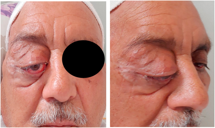
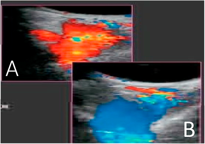
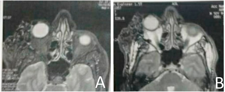
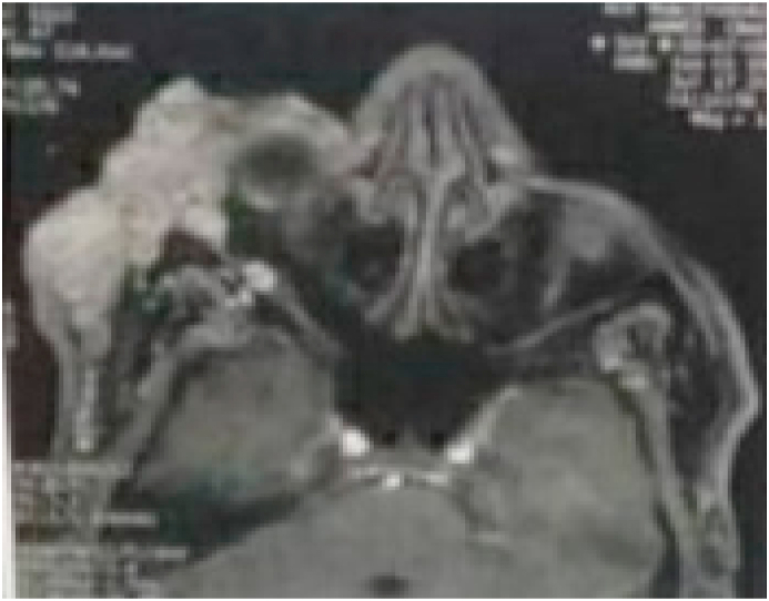
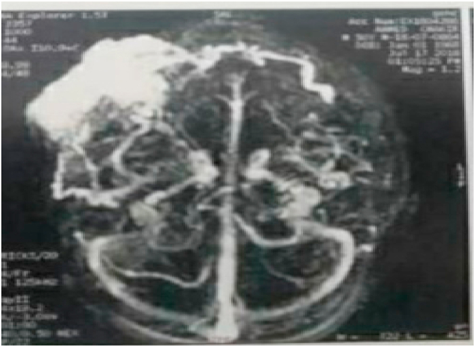
The etiologies of unilateral exophthalmos are multiple, rarely represented by an intra- or extra-conical vascular mass. Orbito-palpebral varixes are rare (2% of orbital masses) and represent a main cause of unilateral intermittent exophthalmos, often of an inflammatory nature. We report a Case of right orbito-palpebral varix in a 65-year-old adult, with no particular history, evolving for 2 years. The ophthalmological examination showed a right palpebral mass, extended to the right external canthus, with palpebral collateral circulation, moderate right ptosis with exophthalmos, non-axial, non-pulsatile, without thrill, painless, without complication, without visual deficit. A vascular mass was suspected and MRI revealed a right orbito-palpebral varix with temporal extension, confirmed by ANGIO-MRI. The latter also allowed to search for a cerebral venous malformation, an encephalocele or a bone defect, associated and also to eliminate differential diagnoses (tumor, arteriovenous fistula …). Color Doppler ultrasound in the proclive position confirmed the diagnosis of orbital varices. A preventive low-dose anticoagulant treatment was started to avoid thrombosis, with therapeutic abstention in the absence of complications. A rigorous monthly follow-up in consultation is ensured. Orbito-palpebral varices are characterized by an extensive posterior intra-orbital character, often during their evolution and imposes a strict surveillance. In Case of complication (thrombosis, hemorrhage, pain, compressive signs of the optic nerve), surgical removal or sclerosis of the varix can be envisaged with disappointing results (recurrence, hemorrhage).
单侧眼球突出的病因多种多样,很少由圆锥内或圆锥外血管性肿物引起。眶睑静脉曲张较为罕见(占眼眶肿物的2%),是单侧间歇性眼球突出的主要原因,通常具有炎症性质。我们报告一例65岁成年人的右侧眶睑静脉曲张病例,无特殊病史,病程2年。眼科检查显示右侧睑部肿物,延伸至右侧外眦,伴有睑部侧支循环,右侧上睑下垂伴眼球突出,非轴向、非搏动性、无震颤、无痛、无并发症、无视力缺损。怀疑为血管性肿物,MRI显示右侧眶睑静脉曲张并向颞侧延伸,磁共振血管造影(ANGIO-MRI)证实了这一诊断。后者还可用于排查是否存在相关的脑静脉畸形、脑膨出或骨缺损,并排除鉴别诊断(肿瘤、动静脉瘘等)。仰卧位彩色多普勒超声证实了眼眶静脉曲张的诊断。开始预防性低剂量抗凝治疗以避免血栓形成,若无并发症则无需进行治疗干预。确保每月进行严格的门诊随访。眶睑静脉曲张的特点是眼眶内后部范围广泛,在其病程中常如此,因此需要严格监测。若出现并发症(血栓形成、出血、疼痛、视神经压迫体征),可考虑手术切除或硬化治疗静脉曲张,但效果往往令人失望(复发、出血)。