Mahmudi Tahereh, Kafieh Raheleh, Rabbani Hossein, Mehri Alireza, Akhlaghi Mohammad-Reza
Department of Medical Physics and Biomedical Engineering, School of Medicine, Tehran University of Medical Sciences, Tehran, Iran.
Medical Image and Signal Processing Research Center, School of Advanced Technologies in Medicine, Isfahan University of Medical Sciences, Isfahan, Iran.
J Med Signals Sens. 2021 Jan 30;11(1):12-23. doi: 10.4103/jmss.JMSS_67_19. eCollection 2021 Jan-Mar.
Asymmetry analysis of retinal layers in right and left eyes can be a valuable tool for early diagnoses of retinal diseases. To determine the limits of the normal interocular asymmetry in retinal layers around macula, thickness measurements are obtained with optical coherence tomography (OCT).
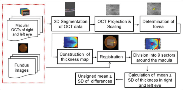
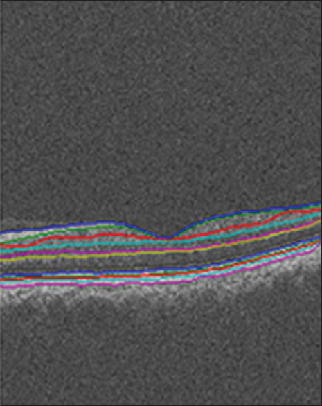
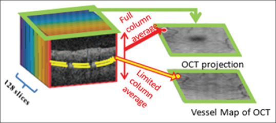
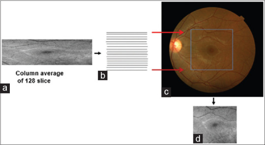
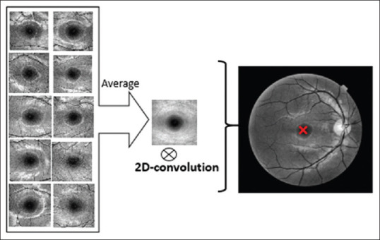
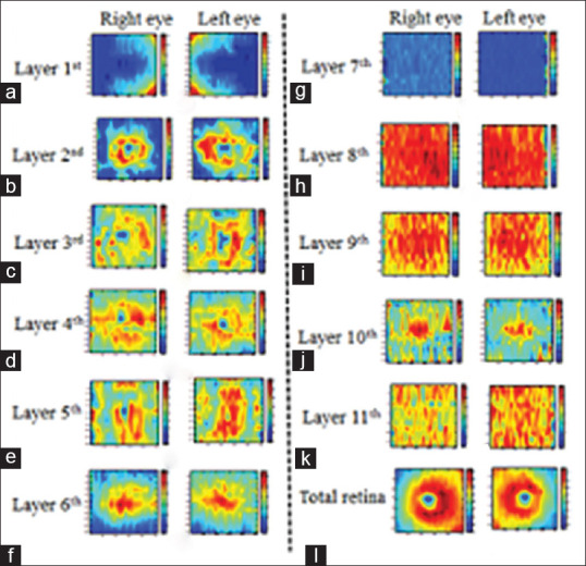
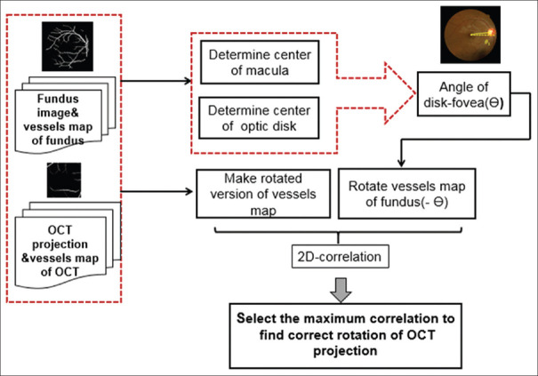
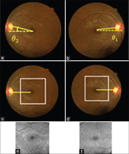
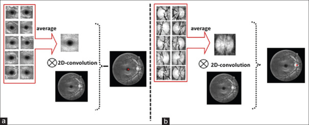
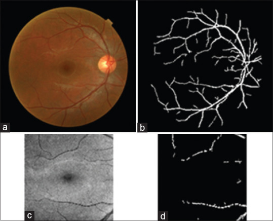
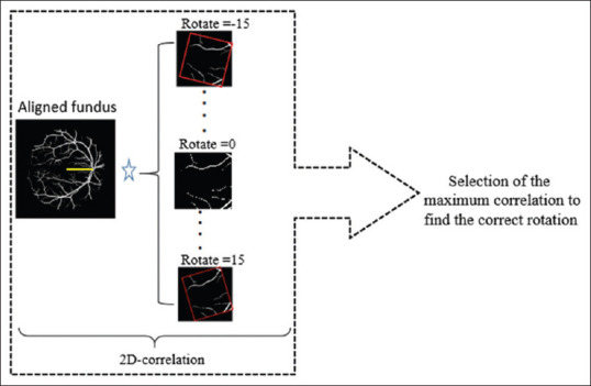
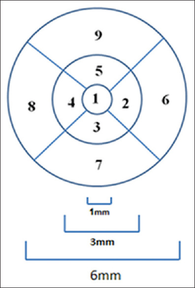
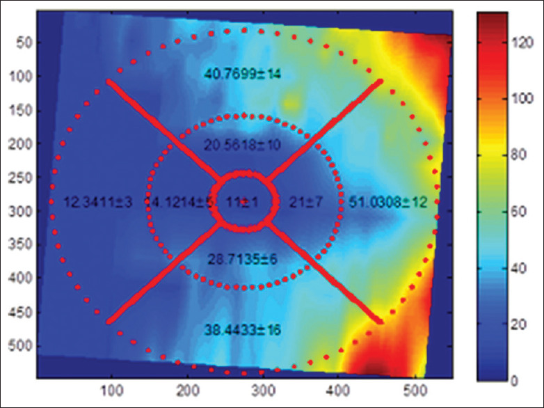
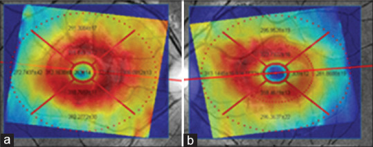
For this purpose, after segmentation of intraretinal layer in threedimensional OCT data and calculating the midmacular point, the TM of each layer is obtained in 9 sectors in concentric circles around the macula. To compare corresponding sectors in the right and left eyes, the TMs of the left and right images are registered by alignment of retinal raphe (i.e. diskfovea axes). Since the retinal raphe of macular OCTs is not calculable due to limited region size, the TMs are registered by first aligning corresponding retinal raphe of fundus images and then registration of the OCTs to aligned fundus images. To analyze the asymmetry in each retinal layer, the mean and standard deviation of thickness in 9 sectors of 11 layers are calculated in 50 normal individuals.
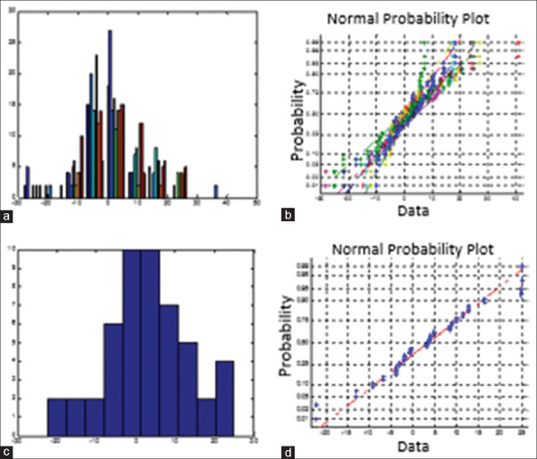
The results demonstrate that some sectors of retinal layers have signifcant asymmetry with < 0.05 in normal population. In this base, the tolerance limits for normal individuals are calculated.
This article shows that normal population does not have identical retinal information in both eyes, and without considering this reality, normal asymmetry in information gathered from both eyes might be interpreted as retinal disorders.
双眼视网膜层的不对称分析可成为视网膜疾病早期诊断的重要工具。为确定黄斑周围视网膜层正常眼间不对称的限度,采用光学相干断层扫描(OCT)进行厚度测量。
为此,在三维OCT数据中分割视网膜内各层并计算黄斑中点后,在围绕黄斑的同心圆的9个扇区中获取每层的厚度均值(TM)。为比较双眼的对应扇区,通过视网膜中缝(即视盘-黄斑轴)对齐来配准左右图像的TM。由于黄斑OCT的视网膜中缝因区域大小有限而无法计算,先通过对齐眼底图像的相应视网膜中缝,然后将OCT配准到对齐的眼底图像来配准TM。为分析各视网膜层的不对称性,计算了50名正常个体11层9个扇区厚度的均值和标准差。
结果表明,视网膜层的一些扇区在正常人群中具有显著不对称性(P<0.05)。在此基础上,计算了正常个体的容许限度。
本文表明正常人群双眼的视网膜信息并不相同,若不考虑这一实际情况,从双眼收集到的信息中的正常不对称性可能会被解释为视网膜疾病。