Biophotonics Research Group, Exploratory Research Center on Life and Living Systems (ExCELLS), National Institutes of Natural Sciences, Higashiyama 5-1, Myodaiji, Okazaki, Aichi 444-8787, Japan.
Division of Biophotonics, National Institute for Physiological Sciences, National Institutes of Natural Sciences, Higashiyama 5-1, Myodaiji, Okazaki, Aichi 444-8787, Japan.
STAR Protoc. 2021 May 12;2(2):100542. doi: 10.1016/j.xpro.2021.100542. eCollection 2021 Jun 18.
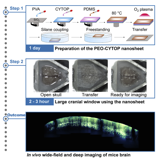
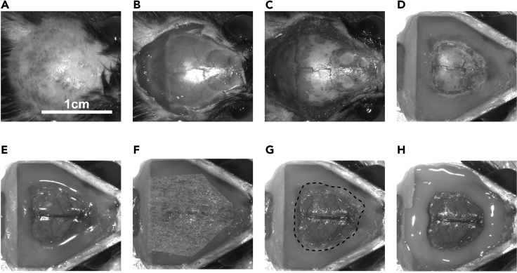
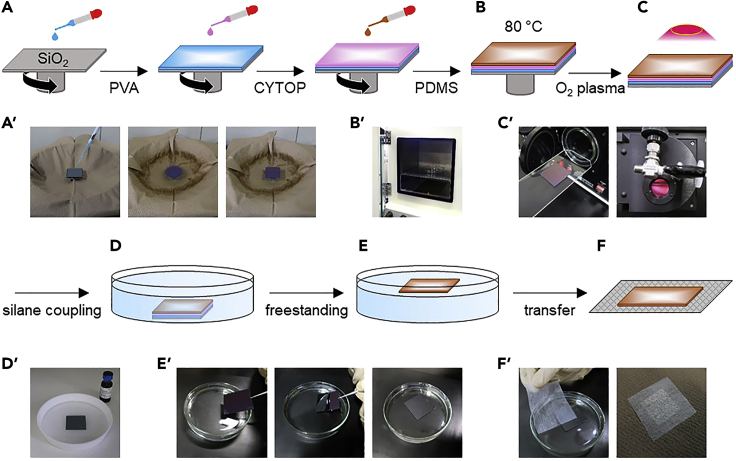
Large-scale optical measurements have revealed the anatomical and functional connectivity among brain regions underlying brain functions. Here, we describe how to construct a cranial window utilizing a polyethylene-oxide-coated CYTOP (PEO-CYTOP) nanosheet that suppresses bleeding on the brain surface of mice. We demonstrate two-photon imaging through the PEO-CYTOP nanosheet at the subcellular resolution in the parietal region of the mouse brain. This protocol improves the surgical procedure and expands the optically observable regions, thereby promoting understanding of brain function. For complete details on the use and execution of this protocol, please refer to Takahashi et al. (2020).
大规模光学测量揭示了大脑功能相关的脑区的解剖和功能连接。在这里,我们描述了如何利用聚氧化乙烯(PEO)包覆的 CYTOP(PEO-CYTOP)纳米片构建颅窗,以抑制小鼠脑表面出血。我们在小鼠大脑顶叶区域展示了通过 PEO-CYTOP 纳米片进行的亚细胞分辨率的双光子成像。该方案改进了手术程序并扩大了可光学观察的区域,从而促进了对大脑功能的理解。如需详细了解本方案的使用和实施,请参考 Takahashi 等人(2020 年)。