Division of Biophotonics, National Institute for Physiological Sciences, National Institutes of Natural Sciences, Higashiyama 5-1, Myodaiji, Okazaki, Aichi, 444-8787, Japan.
Biophotonics Research Group, Exploratory Research Center on Life and Living Systems (ExCELLS), National Institutes of Natural Sciences, Higashiyama 5-1, Myodaiji, Okazaki, Aichi, 444-8787, Japan.
Commun Biol. 2024 Mar 4;7(1):232. doi: 10.1038/s42003-024-05865-8.
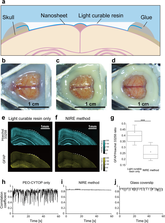
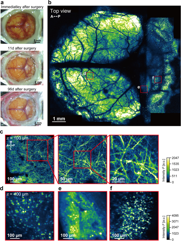
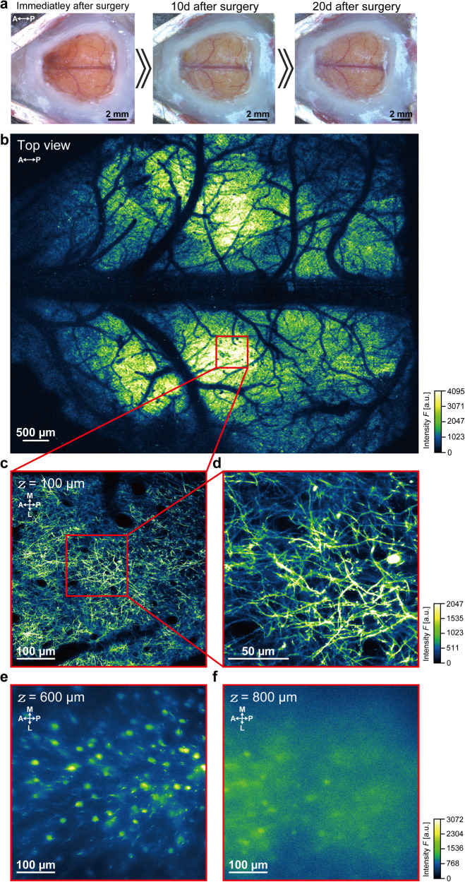
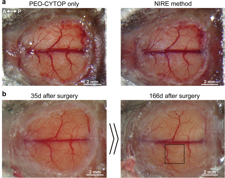
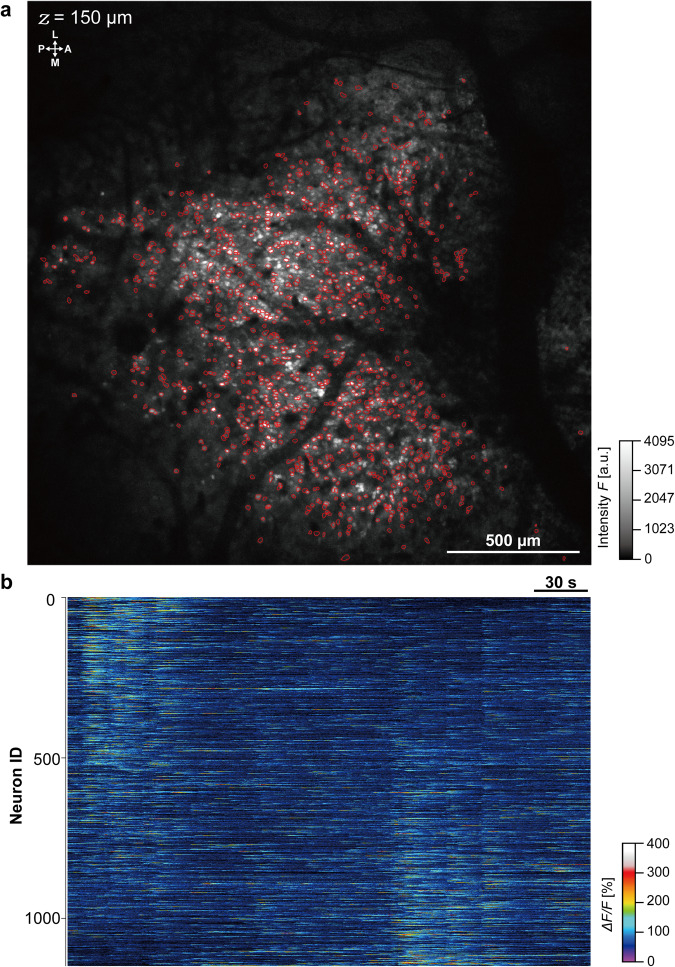
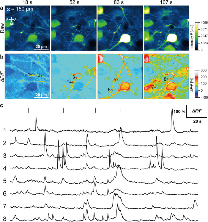
Two-photon microscopy enables in vivo imaging of neuronal activity in mammalian brains at high resolution. However, two-photon imaging tools for stable, long-term, and simultaneous study of multiple brain regions in same mice are lacking. Here, we propose a method to create large cranial windows covering such as the whole parietal cortex and cerebellum in mice using fluoropolymer nanosheets covered with light-curable resin (termed the 'Nanosheet Incorporated into light-curable REsin' or NIRE method). NIRE method can produce cranial windows conforming the curved cortical and cerebellar surfaces, without motion artifacts in awake mice, and maintain transparency for >5 months. In addition, we demonstrate that NIRE method can be used for in vivo two-photon imaging of neuronal ensembles, individual neurons and subcellular structures such as dendritic spines. The NIRE method can facilitate in vivo large-scale analysis of heretofore inaccessible neural processes, such as the neuroplastic changes associated with maturation, learning and neural pathogenesis.
双光子显微镜能够以高分辨率对哺乳动物大脑中的神经元活动进行活体成像。然而,目前还缺乏用于在同一只小鼠中稳定、长期和同时研究多个脑区的双光子成像工具。在这里,我们提出了一种使用覆盖有光固化树脂的氟聚合物纳米片(称为“纳米片掺入光固化树脂”或 NIRE 方法)来创建覆盖小鼠整个顶叶皮层和小脑等区域的大颅窗的方法。NIRE 方法可以产生符合弯曲的皮质和小脑表面的颅窗,在清醒小鼠中没有运动伪影,并保持透明度超过 5 个月。此外,我们证明 NIRE 方法可用于神经元集合、单个神经元和树突棘等亚细胞结构的活体双光子成像。NIRE 方法可以促进对以前无法进入的神经过程进行活体大规模分析,例如与成熟、学习和神经发病机制相关的神经可塑性变化。