Optical Neuroimaging Unit, Okinawa Institute of Science and Technology Graduate University, 1919-1 Tancha, Onna-son, 904-0495 Okinawa, Japan.
STAR Protoc. 2021 Aug 31;2(3):100779. doi: 10.1016/j.xpro.2021.100779. eCollection 2021 Sep 17.
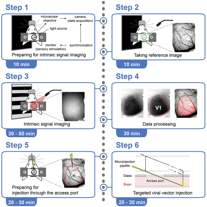
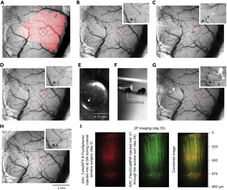
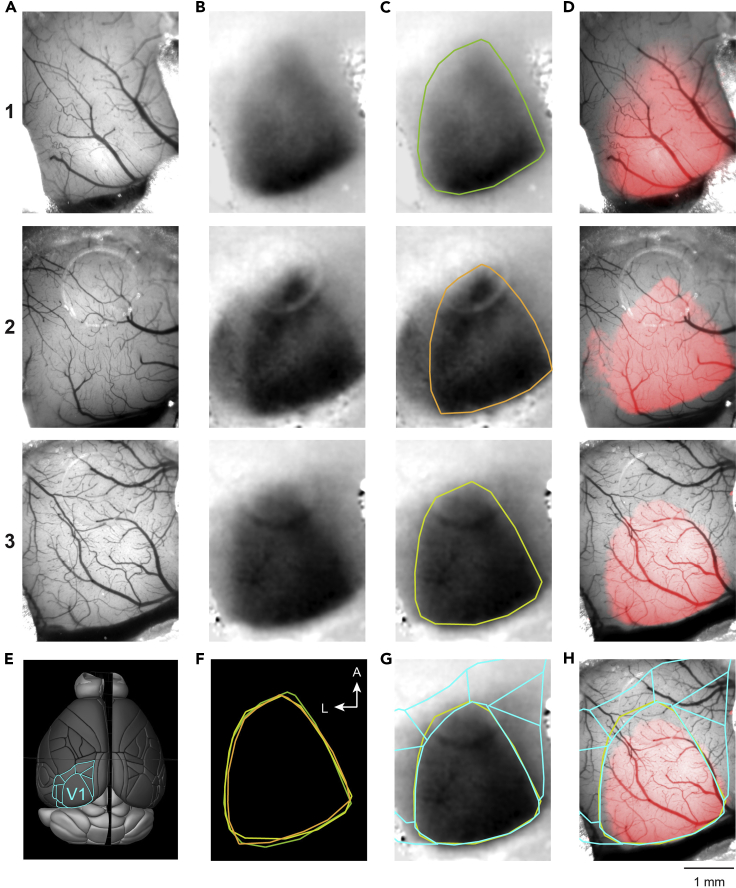
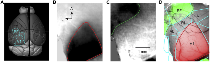
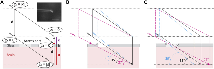
Intrinsic optical signal imaging (ISI) is a hemodynamic response-based technique to map the functional architecture of the cortex. ISI is often used as an auxiliary method to localize cortical areas for targeted electrophysiology, pharmacology, or imaging experiments. Here, we provide a protocol for ISI through a cranial window with an access port to identify the area of the primary visual cortex (V1) in a head-fixed mouse, followed by targeted viral vector injection, which enables subsequent two-photon imaging of V1 layer 6 corticothalamic neurons. For complete details on the use and execution of this protocol, please refer to our paper Augustinaite and Kuhn (2020b).
内源光学信号成像 (ISI) 是一种基于血液动力学反应的技术,用于绘制皮质的功能结构。ISI 通常被用作辅助方法,用于定位皮质区域,以进行靶向电生理学、药理学或成像实验。在这里,我们提供了一种通过带有接入端口的颅窗进行 ISI 的方案,以在头部固定的小鼠中识别初级视觉皮层 (V1) 的区域,然后进行靶向病毒载体注射,这使得随后可以对 V1 层 6 层皮层丘脑神经元进行双光子成像。有关此方案的使用和执行的完整详细信息,请参阅我们的论文 Augustinaite 和 Kuhn (2020b)。