• 文献检索
  • 文档翻译
  • 深度研究
  • 学术资讯
  • Suppr Zotero 插件Zotero 插件
  • 邀请有礼
  • 套餐&价格
  • 历史记录
应用&插件
Suppr Zotero 插件Zotero 插件浏览器插件Mac 客户端Windows 客户端微信小程序
定价
高级版会员购买积分包购买API积分包
服务
文献检索文档翻译深度研究API 文档MCP 服务
关于我们
关于 Suppr公司介绍联系我们用户协议隐私条款
关注我们

Suppr 超能文献

核心技术专利:CN118964589B侵权必究
粤ICP备2023148730 号-1Suppr @ 2026

文献检索

告别复杂PubMed语法,用中文像聊天一样搜索,搜遍4000万医学文献。AI智能推荐,让科研检索更轻松。

立即免费搜索

文件翻译

保留排版,准确专业,支持PDF/Word/PPT等文件格式,支持 12+语言互译。

免费翻译文档

深度研究

AI帮你快速写综述,25分钟生成高质量综述,智能提取关键信息,辅助科研写作。

立即免费体验

无症状疟疾中未成熟网织红细胞和间日疟原虫在脾脏中的蓄积和共定位评估:一项前瞻性人类脾切除术研究。

Evaluation of splenic accumulation and colocalization of immature reticulocytes and Plasmodium vivax in asymptomatic malaria: A prospective human splenectomy study.

机构信息

Global and Tropical Health Division, Menzies School of Health Research and Charles Darwin University, Darwin, Northern Territory, Australia.

Eijkman Institute for Molecular Biology, Jakarta, Indonesia.

出版信息

PLoS Med. 2021 May 26;18(5):e1003632. doi: 10.1371/journal.pmed.1003632. eCollection 2021 May.

DOI:10.1371/journal.pmed.1003632
PMID:34038413
原文链接:https://pmc.ncbi.nlm.nih.gov/articles/PMC8154101/
Abstract

BACKGROUND

A very large biomass of intact asexual-stage malaria parasites accumulates in the spleen of asymptomatic human individuals infected with Plasmodium vivax. The mechanisms underlying this intense tropism are not clear. We hypothesised that immature reticulocytes, in which P. vivax develops, may display high densities in the spleen, thereby providing a niche for parasite survival.

METHODS AND FINDINGS

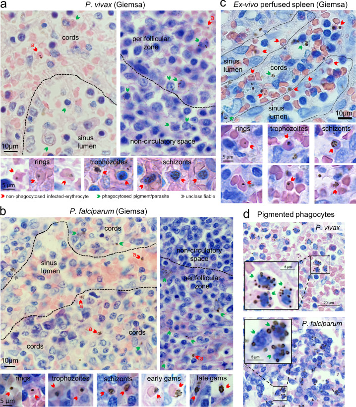
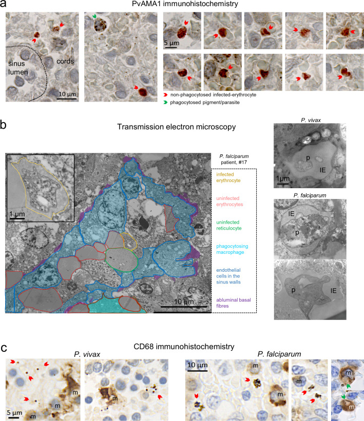
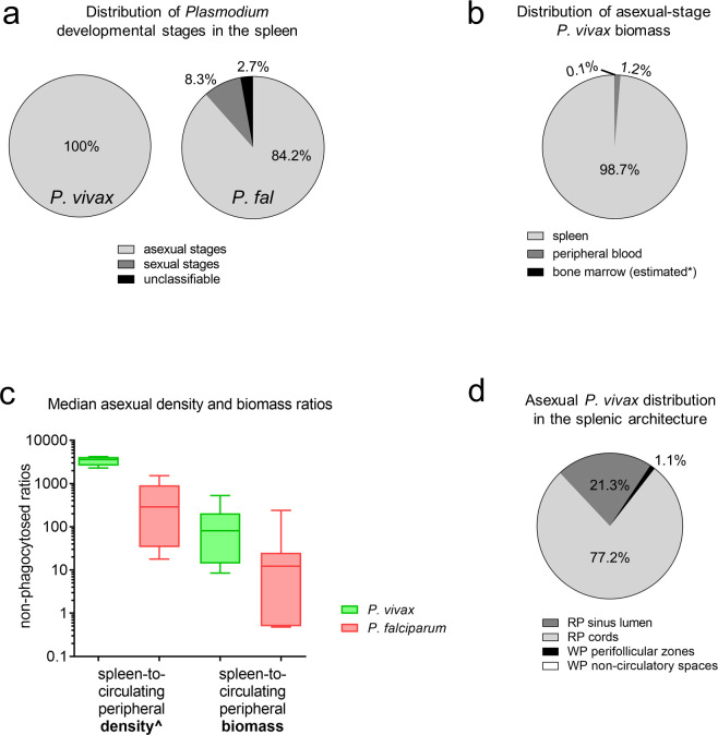
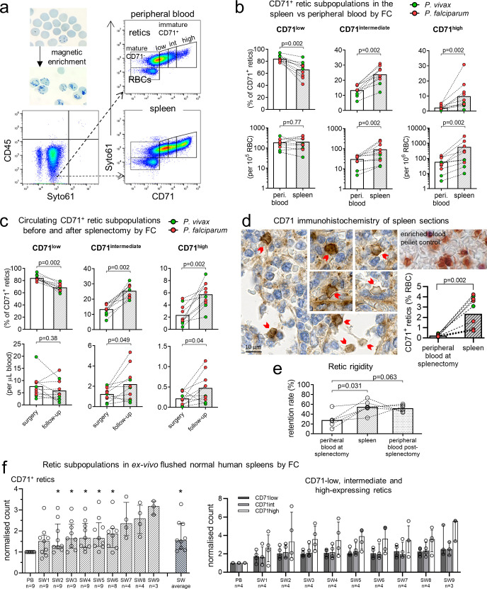
We examined spleen tissue in 22 mostly untreated individuals naturally exposed to P. vivax and Plasmodium falciparum undergoing splenectomy for any clinical indication in malaria-endemic Papua, Indonesia (2015 to 2017). Infection, parasite and immature reticulocyte density, and splenic distribution were analysed by optical microscopy, flow cytometry, and molecular assays. Nine non-endemic control spleens from individuals undergoing spleno-pancreatectomy in France (2017 to 2020) were also examined for reticulocyte densities. There were no exclusion criteria or sample size considerations in both patient cohorts for this demanding approach. In Indonesia, 95.5% (21/22) of splenectomy patients had asymptomatic splenic Plasmodium infection (7 P. vivax, 13 P. falciparum, and 1 mixed infection). Significant splenic accumulation of immature CD71 intermediate- and high-expressing reticulocytes was seen, with concentrations 11 times greater than in peripheral blood. Accordingly, in France, reticulocyte concentrations in the splenic effluent were higher than in peripheral blood. Greater rigidity of reticulocytes in splenic than in peripheral blood, and their higher densities in splenic cords both suggest a mechanical retention process. Asexual-stage P. vivax-infected erythrocytes of all developmental stages accumulated in the spleen, with non-phagocytosed parasite densities 3,590 times (IQR: 2,600 to 4,130) higher than in circulating blood, and median total splenic parasite loads 81 (IQR: 14 to 205) times greater, accounting for 98.7% (IQR: 95.1% to 98.9%) of the estimated total-body P. vivax biomass. More reticulocytes were in contact with sinus lumen endothelial cells in P. vivax- than in P. falciparum-infected spleens. Histological analyses revealed 96% of P. vivax rings/trophozoites and 46% of schizonts colocalised with 92% of immature reticulocytes in the cords and sinus lumens of the red pulp. Larger splenic cohort studies and similar investigations in untreated symptomatic malaria are warranted.

CONCLUSIONS

Immature CD71+ reticulocytes and splenic P. vivax-infected erythrocytes of all asexual stages accumulate in the same splenic compartments, suggesting the existence of a cryptic endosplenic lifecycle in chronic P. vivax infection. Findings provide insight into P. vivax-specific adaptions that have evolved to maximise survival and replication in the spleen.

摘要

背景

无症状感染间日疟原虫的人体脾脏中会积累大量完整的无性生殖阶段疟原虫。这种强烈的嗜脾性的机制尚不清楚。我们假设幼稚网织红细胞(疟原虫在其中发育)在脾脏中可能显示出高密度,从而为寄生虫的存活提供了一个小生境。

方法和发现

我们在印度尼西亚巴布亚(2015 年至 2017 年)进行了任何临床指征的脾切除术的 22 名主要未经治疗的自然暴露于间日疟原虫和恶性疟原虫的个体的脾脏组织中进行了检查。通过光学显微镜、流式细胞术和分子检测分析了感染、寄生虫和幼稚网织红细胞密度以及脾脏分布。法国在 2017 年至 2020 年进行脾胰切除术的 9 名非流行地区对照脾脏也进行了网织红细胞密度检查。对于这种要求很高的方法,两组患者队列都没有排除标准或样本量考虑。在印度尼西亚,95.5%(21/22)的脾切除术患者存在无症状的脾疟原虫感染(7 例间日疟原虫、13 例恶性疟原虫和 1 例混合感染)。在脾脏中可以看到显著的幼稚 CD71 中表达和高表达网织红细胞的积聚,浓度比外周血高 11 倍。相应地,在法国,脾流出物中的网织红细胞浓度高于外周血。与外周血相比,脾内网织红细胞的刚性更大,脾内网织红细胞的密度更高,这都表明存在机械滞留过程。所有发育阶段的间日疟原虫感染的有性期红细胞都积聚在脾脏中,未被吞噬的寄生虫密度比循环血液高 3590 倍(IQR:2600 至 4130),总脾脏寄生虫负荷中位数高 81 倍(IQR:14 至 205),占估计的间日疟原虫总生物量的 98.7%(IQR:95.1%至 98.9%)。与恶性疟原虫感染的脾脏相比,间日疟原虫感染的脾脏中有更多的网织红细胞与窦腔内皮细胞接触。组织学分析显示,96%的间日疟原虫环/滋养体和 46%的裂殖体与红髓窦腔和索中的 92%幼稚网织红细胞共定位。需要进行更大规模的脾脏队列研究和对未经治疗的有症状疟疾进行类似的研究。

结论

幼稚的 CD71+网织红细胞和所有无性生殖阶段的间日疟原虫感染的红细胞在相同的脾脏隔室中积聚,这表明在慢性间日疟原虫感染中存在隐匿性脾内生命周期。这些发现为间日疟原虫特有的适应提供了深入了解,这些适应已经进化到最大限度地提高了在脾脏中的生存和复制能力。

https://cdn.ncbi.nlm.nih.gov/pmc/blobs/d537/8154101/427e2a2172f0/pmed.1003632.g005.jpg
https://cdn.ncbi.nlm.nih.gov/pmc/blobs/d537/8154101/2367a17c4e75/pmed.1003632.g001.jpg
https://cdn.ncbi.nlm.nih.gov/pmc/blobs/d537/8154101/071be9f9676a/pmed.1003632.g002.jpg
https://cdn.ncbi.nlm.nih.gov/pmc/blobs/d537/8154101/924e569df43f/pmed.1003632.g003.jpg
https://cdn.ncbi.nlm.nih.gov/pmc/blobs/d537/8154101/8065a5538c46/pmed.1003632.g004.jpg
https://cdn.ncbi.nlm.nih.gov/pmc/blobs/d537/8154101/427e2a2172f0/pmed.1003632.g005.jpg

相似文献

1
Evaluation of splenic accumulation and colocalization of immature reticulocytes and Plasmodium vivax in asymptomatic malaria: A prospective human splenectomy study.无症状疟疾中未成熟网织红细胞和间日疟原虫在脾脏中的蓄积和共定位评估:一项前瞻性人类脾切除术研究。
PLoS Med. 2021 May 26;18(5):e1003632. doi: 10.1371/journal.pmed.1003632. eCollection 2021 May.
2
Reticulocyte-prone malaria parasites predominantly invade CD71hi immature cells: implications for the development of an in vitro culture for Plasmodium vivax.易成网织红细胞疟疾寄生虫主要侵袭 CD71hi 幼稚细胞:对开发体外培养间日疟原虫的影响。
Malar J. 2013 Dec 1;12:434. doi: 10.1186/1475-2875-12-434.
3
Positron emission tomography and magnetic resonance imaging in experimental human malaria to identify organ-specific changes in morphology and glucose metabolism: A prospective cohort study.正电子发射断层扫描和磁共振成像在实验性人类疟疾中识别形态和葡萄糖代谢的器官特异性变化:一项前瞻性队列研究。
PLoS Med. 2021 May 26;18(5):e1003567. doi: 10.1371/journal.pmed.1003567. eCollection 2021 May.
4
DARC extracellular domain remodeling in maturating reticulocytes explains tropism.成熟网织红细胞中DARC细胞外结构域重塑解释了嗜性。
Blood. 2017 Sep 21;130(12):1441-1444. doi: 10.1182/blood-2017-03-774364. Epub 2017 Jul 28.
5
Reticulocyte Preference and Stage Development of Plasmodium vivax Isolates.间日疟原虫分离株的网织红细胞偏好性与阶段发育
J Infect Dis. 2016 Oct 1;214(7):1081-4. doi: 10.1093/infdis/jiw303. Epub 2016 Jul 17.
6
The biology and pathogenesis of vivax malaria.间日疟原虫的生物学和发病机制。
Trends Parasitol. 2024 Jul;40(7):573-590. doi: 10.1016/j.pt.2024.04.015. Epub 2024 May 15.
7
Hidden Biomass of Intact Malaria Parasites in the Human Spleen.人类脾脏中完整疟原虫的隐藏生物量。
N Engl J Med. 2021 May 27;384(21):2067-2069. doi: 10.1056/NEJMc2023884.
8
Plasmodium vivax: restricted tropism and rapid remodeling of CD71-positive reticulocytes.间日疟原虫:CD71 阳性网织红细胞的嗜性受限与快速重塑
Blood. 2015 Feb 19;125(8):1314-24. doi: 10.1182/blood-2014-08-596015. Epub 2014 Nov 20.
9
Plasmodium vivax infection compromises reticulocyte stability.间日疟原虫感染会损害网织红细胞的稳定性。
Nat Commun. 2021 Mar 12;12(1):1629. doi: 10.1038/s41467-021-21886-x.
10
Preserved dendritic cell HLA-DR expression and reduced regulatory T cell activation in asymptomatic Plasmodium falciparum and P. vivax infection.无症状恶性疟原虫和间日疟原虫感染中树突状细胞HLA-DR表达得以保留且调节性T细胞活化降低
Infect Immun. 2015 Aug;83(8):3224-32. doi: 10.1128/IAI.00226-15. Epub 2015 Jun 1.

引用本文的文献

1
Systems approach identifies monocyte imbalance in symptomatic and asymptomatic P. vivax malaria.系统方法识别有症状和无症状间日疟原虫疟疾中的单核细胞失衡。
Mol Syst Biol. 2025 Aug 19. doi: 10.1038/s44320-025-00135-z.
2
Preliminary assessment of serum endocan as a biomarker of disease severity in Plasmodium falciparum and Plasmodium vivax malaria.血清内配蛋白作为恶性疟原虫和间日疟原虫疟疾疾病严重程度生物标志物的初步评估。
Malar J. 2025 Jul 18;24(1):236. doi: 10.1186/s12936-025-05483-7.
3
Reversible host cell surface remodelling limits immune recognition and maximizes survival of Plasmodium falciparum gametocytes.

本文引用的文献

1
Hidden Biomass of Intact Malaria Parasites in the Human Spleen.人类脾脏中完整疟原虫的隐藏生物量。
N Engl J Med. 2021 May 27;384(21):2067-2069. doi: 10.1056/NEJMc2023884.
2
Positron emission tomography and magnetic resonance imaging in experimental human malaria to identify organ-specific changes in morphology and glucose metabolism: A prospective cohort study.正电子发射断层扫描和磁共振成像在实验性人类疟疾中识别形态和葡萄糖代谢的器官特异性变化:一项前瞻性队列研究。
PLoS Med. 2021 May 26;18(5):e1003567. doi: 10.1371/journal.pmed.1003567. eCollection 2021 May.
3
Rapid clearance of storage-induced microerythrocytes alters transfusion recovery.
可逆的宿主细胞表面重塑限制了免疫识别并使恶性疟原虫配子体的存活率最大化。
PLoS Pathog. 2025 May 12;21(5):e1013110. doi: 10.1371/journal.ppat.1013110. eCollection 2025 May.
4
Leveraging microphysiological systems to expedite understanding of host-parasite interactions.利用微生理系统加快对宿主-寄生虫相互作用的理解。
PLoS Pathog. 2025 Apr 24;21(4):e1013088. doi: 10.1371/journal.ppat.1013088. eCollection 2025 Apr.
5
Malaria.疟疾
N Engl J Med. 2025 Apr 3;392(13):1320-1333. doi: 10.1056/NEJMra2405313.
6
Asymptomatic vivax malaria is associated with an IFN-γ-program on adaptive immunity.无症状间日疟与适应性免疫上的一个干扰素-γ程序相关。
J Transl Med. 2025 Mar 19;23(1):351. doi: 10.1186/s12967-025-06353-1.
7
Extracellular vesicles in malaria: proteomics insights, and studies indicate the need for transitioning to natural human infections.疟疾中的细胞外囊泡:蛋白质组学见解,以及研究表明需要转向天然人类感染。
mBio. 2025 Mar 12;16(3):e0230424. doi: 10.1128/mbio.02304-24. Epub 2025 Jan 27.
8
Potent AMA1-specific human monoclonal antibody against Plasmodium vivax Pre-erythrocytic and Blood Stages.针对间日疟原虫前体红细胞期和血液期的强效AMA1特异性人单克隆抗体。
Nat Commun. 2024 Dec 4;15(1):10556. doi: 10.1038/s41467-024-53848-4.
9
Clearance of pathogenic erythrocytes is maintained despite spleen dysfunction in children with sickle cell disease.尽管患有镰状细胞病的儿童脾脏功能失调,但仍能清除致病的红细胞。
Am J Hematol. 2024 Dec;99(12):2267-2278. doi: 10.1002/ajh.27481. Epub 2024 Sep 17.
10
Lineage-informative microhaplotypes for recurrence classification and spatio-temporal surveillance of Plasmodium vivax malaria parasites.用于间日疟原虫疟疾寄生虫复发分类和时空监测的谱系信息微单倍型
Nat Commun. 2024 Aug 8;15(1):6757. doi: 10.1038/s41467-024-51015-3.
储存诱导的小红细胞快速清除改变了输血恢复。
Blood. 2021 Apr 29;137(17):2285-2298. doi: 10.1182/blood.2020008563.
4
Morphological and Transcriptional Changes in Human Bone Marrow During Natural Plasmodium vivax Malaria Infections.人体骨髓在自然感染间日疟原虫过程中的形态和转录变化。
J Infect Dis. 2022 Apr 1;225(7):1274-1283. doi: 10.1093/infdis/jiaa177.
5
Plasma-derived extracellular vesicles from Plasmodium vivax patients signal spleen fibroblasts via NF-kB facilitating parasite cytoadherence.来自间日疟原虫患者的血浆衍生细胞外囊泡通过 NF-κB 信号转导作用于脾成纤维细胞,促进寄生虫细胞黏附。
Nat Commun. 2020 Jun 2;11(1):2761. doi: 10.1038/s41467-020-16337-y.
6
spleen-dependent genes encode antigens associated with cytoadhesion and clinical protection.脾依赖基因编码与细胞黏附和临床保护相关的抗原。
Proc Natl Acad Sci U S A. 2020 Jun 9;117(23):13056-13065. doi: 10.1073/pnas.1920596117. Epub 2020 May 21.
7
Plasmodium vivax in the Era of the Shrinking P. falciparum Map.《在恶性疟原虫地图不断缩小的时代中的间日疟原虫》
Trends Parasitol. 2020 Jun;36(6):560-570. doi: 10.1016/j.pt.2020.03.009. Epub 2020 Apr 22.
8
Analysis of Global Research on Malaria and .疟疾及. 的全球研究分析。
Int J Environ Res Public Health. 2019 May 31;16(11):1928. doi: 10.3390/ijerph16111928.
9
Extramedullary hematopoiesis in the absence of myeloproliferative neoplasm: Mayo Clinic case series of 309 patients.无骨髓增殖性肿瘤情况下的髓外造血:梅奥诊所309例患者病例系列
Blood Cancer J. 2018 Nov 19;8(12):119. doi: 10.1038/s41408-018-0156-6.
10
Niche-induced extramedullary hematopoiesis in the spleen is regulated by the transcription factor Tlx1.龛诱导的脾脏髓外造血由转录因子 Tlx1 调节。
Sci Rep. 2018 May 29;8(1):8308. doi: 10.1038/s41598-018-26693-x.