Sichuan Key Laboratory of Medical Imaging, Department of Radiology, Affiliated Hospital of North Sichuan Medical College, 63# Wenhua Road, 637000, Nanchong, Sichuan, China.
Sichuan Key Laboratory of Medical Imaging, North Sichuan Medical College, Nanchong, Sichuan, China.
Cancer Imaging. 2021 May 26;21(1):38. doi: 10.1186/s40644-021-00407-5.
Early recurrence of oesophageal squamous cell carcinoma (SCC) is defined as recurrence after surgery within 1 year, and appears as local recurrence, distant recurrence, and lymph node positive and disseminated recurrence. Contrast-enhanced computed tomography (CECT) is recommended for diagnosis of primary tumor and initial staging of oesophageal SCC, but it cannot be used to predict early recurrence. It is reported that radiomics can help predict preoperative stages of oesophageal SCC, lymph node metastasis before operation, and 3-year overall survival of oesophageal SCC patients following chemoradiotherapy by extracting high-throughput quantitative features from CT images. This study aimed to develop models based on CT radiomics and clinical features of oesophageal SCC to predict early recurrence of locally advanced cancer.
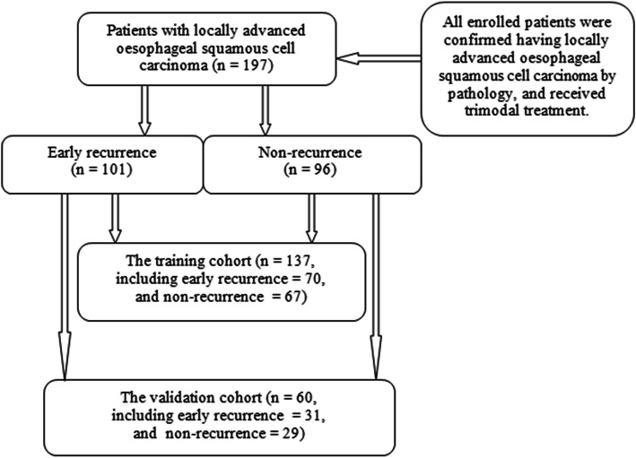
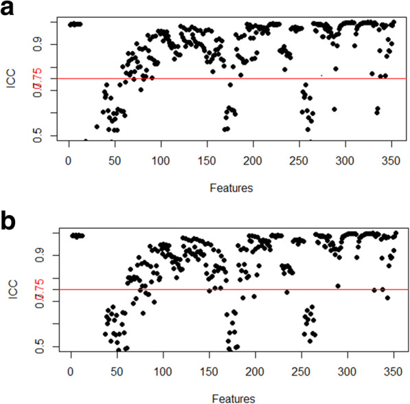
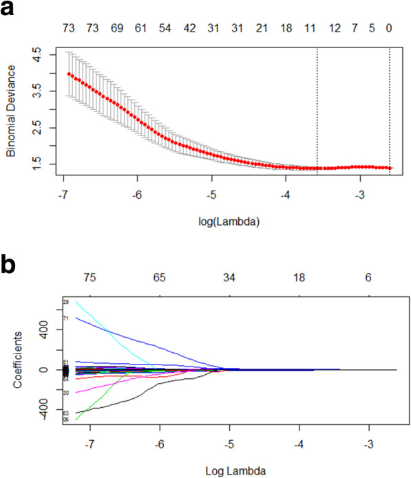
We collected electronic medical records and image data of 197 patients with confirmed locally advanced oesophageal SCC. These patients were randomly allocated to 137 patients in the training cohort and 60 in the test cohort. 352 radiomics features were extracted by delineating region-of-interest (ROI) around the lesion on CECT images and clinical signature was generated by medical records. The radiomics model, clinical model, the combined model of radiomics and clinical features were developed by radiomics features and/or clinical characteristics. Predicting performance of the three models was assessed with area under receiver operating characteristic curve (AUC), accuracy and F-1 score.
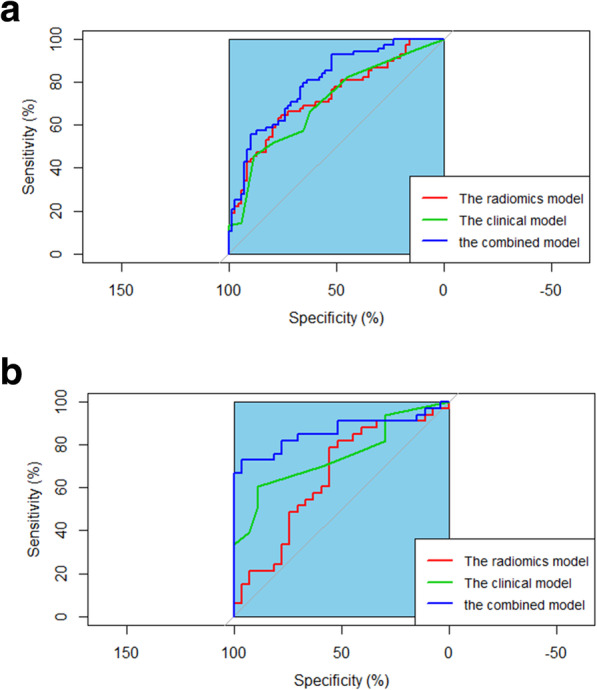
Eleven radiomics features and/or six clinical signatures were selected to build prediction models related to recurrence of locally advanced oesophageal SCC after trimodal therapy. The AUC of integration of radiomics and clinical models was better than that of radiomics or clinical model for the training cohort (0.821 versus 0.754 or 0.679, respectively) and for the validation cohort (0.809 versus 0.646 or 0.658, respectively). Integrated model of radiomics and clinical features showed good performance in predicting early recurrence of locally advanced oesophageal SCC for both the training and validation cohorts (accuracy = 0.730 and 0.733, and F-1score = 0.730 and 0.778, respectively).
The integrated model of CECT radiomics and clinical features may be a potential imaging biomarker to predict early recurrence of locally advanced oesophageal SCC after trimodal therapy.
食管鳞状细胞癌(SCC)的早期复发定义为手术后 1 年内的复发,表现为局部复发、远处复发以及淋巴结阳性和播散性复发。增强计算机断层扫描(CECT)推荐用于诊断原发性肿瘤和食管 SCC 的初始分期,但不能用于预测早期复发。据报道,通过从 CT 图像中提取高通量定量特征,放射组学可以帮助预测食管 SCC 的术前分期、手术前的淋巴结转移以及接受放化疗的食管 SCC 患者的 3 年总生存率。本研究旨在建立基于 CT 放射组学和食管 SCC 临床特征的模型,以预测局部晚期癌症的早期复发。
我们收集了 197 例经证实的局部晚期食管 SCC 患者的电子病历和图像数据。这些患者被随机分配到训练队列的 137 例和测试队列的 60 例。通过在 CECT 图像上勾画病变的感兴趣区域(ROI)提取 352 个放射组学特征,并通过病历生成临床特征。通过放射组学特征和/或临床特征构建放射组学模型、临床模型和放射组学与临床特征的联合模型。通过受试者工作特征曲线(ROC)下面积(AUC)、准确性和 F-1 评分评估三种模型的预测性能。
选择 11 个放射组学特征和/或 6 个临床特征来建立与局部晚期食管 SCC 经三联疗法治疗后复发相关的预测模型。放射组学与临床模型的整合 AUC 优于放射组学或临床模型在训练队列(0.821 对 0.754 或 0.679)和验证队列(0.809 对 0.646 或 0.658)中的表现。放射组学与临床特征的综合模型在训练和验证队列中均能很好地预测局部晚期食管 SCC 的早期复发(准确性分别为 0.730 和 0.733,F-1 评分分别为 0.730 和 0.778)。
CECT 放射组学和临床特征的综合模型可能是预测局部晚期食管 SCC 经三联疗法治疗后早期复发的潜在影像学生物标志物。