Horsley Kristen T, Olby Natasha J, Mitchell Mark A, Aulakh Karanvir S, Gines J Alberto
Department of Veterinary Clinical Sciences, School of Veterinary Medicine, Louisiana State University, Baton Rouge, LA, United States.
College of Veterinary Medicine, North Carolina State University, Raleigh, NC, United States.
Front Vet Sci. 2021 May 10;8:664150. doi: 10.3389/fvets.2021.664150. eCollection 2021.
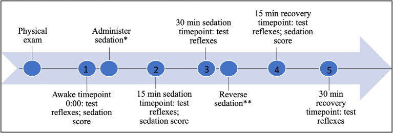
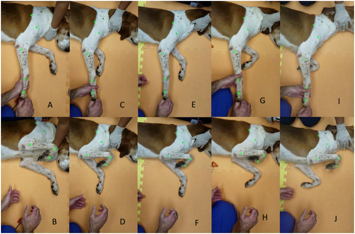
Pain, temperament, fear, and anxiety can prevent safe and accurate evaluation of common neurologic reflexes in dogs. When sedation is used it is unknown how the neurological examination, and specifically patellar and withdrawal reflexes are affected, and, if present, how long any effect might last. The purpose of this study is to investigate the effect of sedation on the evaluation of select common limb spinal reflexes in healthy dogs. Fourteen healthy dogs with normal neurologic exams were included. After placing joint landmarks, patellar reflex and pelvic and thoracic limb withdrawal reflexes were tested. Joint angles were measured, obtaining reflex angle endpoints, change in angle, and change in time to reflex completion. These measurements were recorded at different time points: prior to sedation (awake timepoint), 15 and 30 min following administration of standardized sedation protocol of dexmedetomidine and butorphanol, and 15 and 30 min following administration of a standardized reversal agent, atipamazole. For patellar reflex, the stifle end angle increased from 91.5 to 108.55 degrees ( < 0.0001) 15 min following sedation, and remained increased at 104.5 degrees ( < 0.0001) 30 min following sedation. Stifle change in angle increased from 9.6 to 24.4 degrees ( < 0.0001) 15 min following sedation, and remained increased at 20.85 degrees ( < 0.0001) and 11 degrees ( = 0.012) at 30 min sedation and 15 min reversal. Tarsal joint in pelvic withdrawal and elbow in thoracic withdrawal reflexes did not differ in at any timepoint of sedation or reversal when compared with the awake timepoint, for end angle or change in angle. The increases in end angle and change in angle for patellar reflex generated a change in time for patellar reflex from 0.12 s (awake) to 0.129 s (15 min sedation) which was statistically significant ( = 0.041). Change in time did not differ for pelvic withdrawal or thoracic withdrawal. Reflexes were elicited in all dogs under sedation. Sedation does not affect the evaluation of the withdrawal reflex on any limb but improves the visualization of the patellar reflex in this group of neurologically normal dogs.
疼痛、性情、恐惧和焦虑会妨碍对犬类常见神经反射进行安全准确的评估。使用镇静剂时,尚不清楚神经学检查,特别是髌反射和退缩反射会受到怎样的影响,以及如果有影响的话,这种影响可能会持续多久。本研究的目的是调查镇静对健康犬选定的常见肢体脊髓反射评估的影响。纳入了14只神经学检查正常的健康犬。在确定关节标志点后,测试髌反射以及骨盆和胸肢的退缩反射。测量关节角度,获取反射角终点、角度变化以及反射完成时间的变化。这些测量在不同时间点记录:镇静前(清醒时间点)、给予右美托咪定和布托啡诺标准化镇静方案后15分钟和30分钟,以及给予标准化逆转剂阿替美唑后15分钟和30分钟。对于髌反射,镇静后15分钟时, stifle终末角度从91.5度增加到108.55度(<0.0001),镇静后30分钟时保持在104.5度增加(<0.0001)。镇静后15分钟时, stifle角度变化从9.6度增加到24.4度(<0.0001),镇静30分钟和逆转15分钟时分别保持在20.85度增加(<0.0001)和11度增加(=0.012)。与清醒时间点相比,在镇静或逆转的任何时间点,骨盆退缩反射中的跗关节和胸肢退缩反射中的肘关节在终末角度或角度变化方面均无差异。髌反射终末角度和角度变化的增加导致髌反射时间从0.12秒(清醒)变为0.129秒(镇静15分钟),具有统计学意义(=0.041)。骨盆退缩或胸肢退缩的时间变化无差异。所有犬在镇静状态下均可引出反射。在这组神经学正常的犬中,镇静不影响对任何肢体退缩反射的评估,但改善了髌反射的观察效果。