Wang Dinghui, Xiong Tianhua, Yu Wenlong, Liu Bin, Wang Jing, Xiao Kaihu, She Qiang
Department of Cardiology, The Second Affiliated Hospital of Chongqing Medical University, Chongqing, China.
Front Genet. 2021 May 11;12:650213. doi: 10.3389/fgene.2021.650213. eCollection 2021.
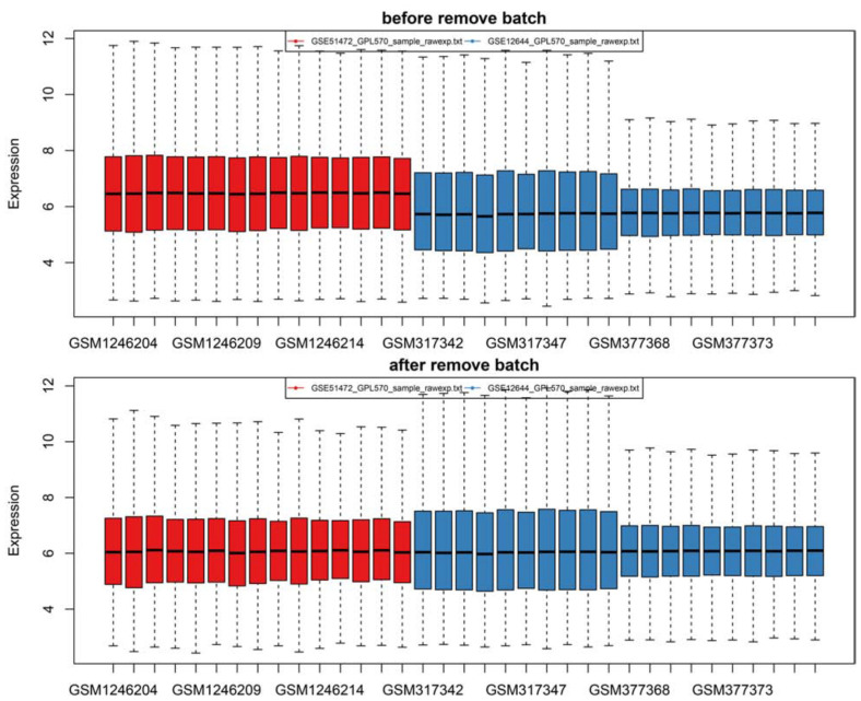
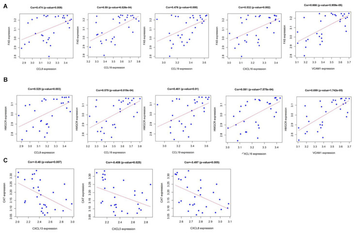
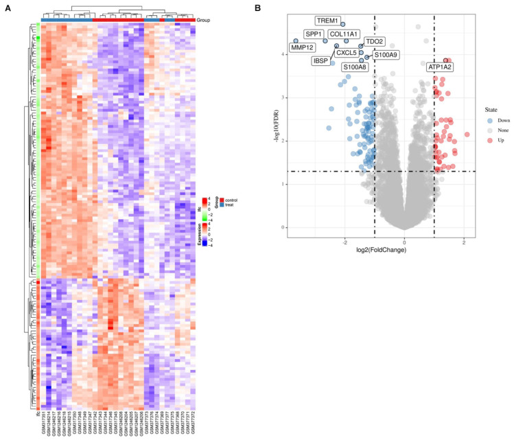
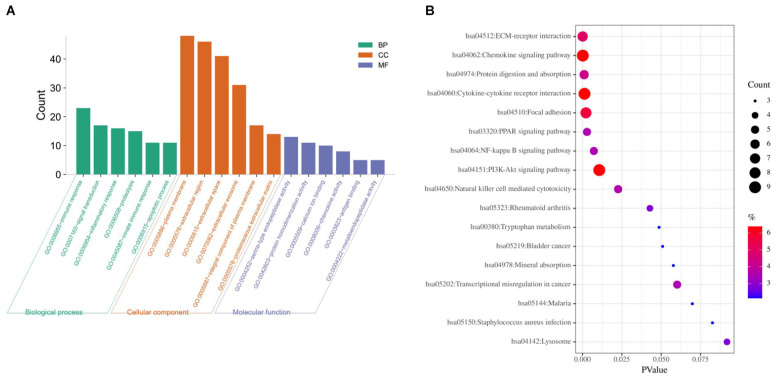
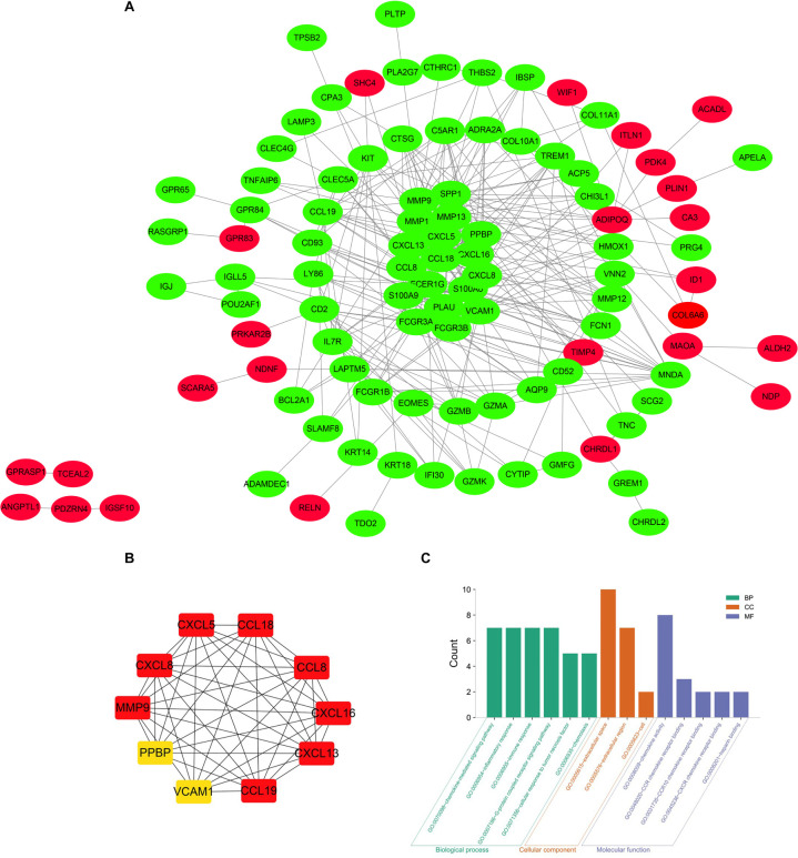
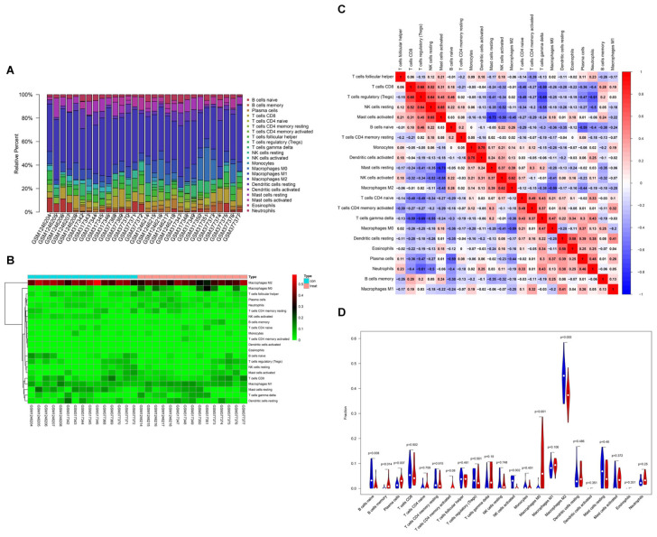
Valvular heart disease is obtaining growing attention in the cardiovascular field and it is believed that calcific aortic valve disease (CAVD) is the most common valvular heart disease (VHD) in the world. CAVD does not have a fully effective treatment to delay its progression and the specific molecular mechanism of aortic valve calcification remains unclear. We obtained the gene expression datasets GSE12644 and GSE51472 from the public comprehensive free database GEO. Then, a series of bioinformatics methods, such as GO and KEGG analysis, STING online tool, Cytoscape software, were used to identify differentially expressed genes in CAVD and healthy controls, construct a PPI network, and then identify key genes. In addition, immune infiltration analysis was used via CIBERSORT to observe the expression of various immune cells in CAVD. A total of 144 differential expression genes were identified in the CAVD samples in comparison with the control samples, including 49 up-regulated genes and 95 down-regulated genes. GO analysis of DEGs were most observably enriched in the immune response, signal transduction, inflammatory response, proteolysis, innate immune response, and apoptotic process. The KEGG analysis revealed that the enrichment of DEGs in CAVD were remarkably observed in the chemokine signaling pathway, cytokine-cytokine receptor interaction, and PI3K-Akt signaling pathway. Chemokines CXCL13, CCL19, CCL8, CXCL8, CXCL16, MMP9, CCL18, CXCL5, VCAM1, and PPBP were identified as the hub genes of CAVD. It was macrophages that accounted for the maximal proportion among these immune cells. The expression of macrophages M0, B cells memory, and Plasma cells were higher in the CAVD valves than in healthy valves, however, the expression of B cells naïve, NK cells activated, and macrophages M2 were lower. We detected that chemokines CXCL13, CXCL8, CXCL16, and CXCL5, and CCL19, CCL8, and CCL18 are the most important markers of aortic valve disease. The regulatory macrophages M0, plasma cells, B cells memory, B cells naïve, NK cells activated, and macrophages M2 are probably related to the occurrence and the advancement of aortic valve stenosis. These identified chemokines and these immune cells may interact with a subtle adjustment relationship in the development of calcification in CAVD.
心脏瓣膜病在心血管领域日益受到关注,据信钙化性主动脉瓣疾病(CAVD)是全球最常见的心脏瓣膜病(VHD)。CAVD尚无完全有效的治疗方法来延缓其进展,主动脉瓣钙化的具体分子机制仍不清楚。我们从公共综合免费数据库GEO中获取了基因表达数据集GSE12644和GSE51472。然后,使用一系列生物信息学方法,如GO和KEGG分析、STING在线工具、Cytoscape软件,来识别CAVD和健康对照中的差异表达基因,构建蛋白质-蛋白质相互作用(PPI)网络,进而识别关键基因。此外,通过CIBERSORT进行免疫浸润分析,以观察CAVD中各种免疫细胞的表达。与对照样本相比,在CAVD样本中总共鉴定出144个差异表达基因,包括49个上调基因和95个下调基因。对差异表达基因的GO分析最显著地富集于免疫应答、信号转导、炎症应答、蛋白水解、固有免疫应答和凋亡过程。KEGG分析显示,CAVD中差异表达基因在趋化因子信号通路、细胞因子-细胞因子受体相互作用和PI3K-Akt信号通路中显著富集。趋化因子CXCL13、CCL19、CCL8、CXCL8、CXCL16、MMP9、CCL18、CXCL5、VCAM1和PPBP被鉴定为CAVD的枢纽基因。在这些免疫细胞中,巨噬细胞占比最大。CAVD瓣膜中巨噬细胞M0、记忆B细胞和浆细胞的表达高于健康瓣膜,然而,幼稚B细胞、活化NK细胞和巨噬细胞M2的表达较低。我们检测到趋化因子CXCL13、CXCL8、CXCL16和CXCL5以及CCL19、CCL8和CCL18是主动脉瓣疾病的最重要标志物。调节性巨噬细胞M0、浆细胞、记忆B细胞、幼稚B细胞、活化NK细胞和巨噬细胞M2可能与主动脉瓣狭窄的发生和进展有关。这些鉴定出的趋化因子和这些免疫细胞可能在CAVD钙化发展过程中以微妙的调节关系相互作用。