Ryota Hironori, Ishida Mitsuaki, Ebisu Yusuke, Yanagimoto Hiroaki, Yamamoto Tomohisa, Kosaka Hisashi, Hirooka Satoshi, Yamaki So, Kotsuka Masaya, Matsui Yoichi, Tsuta Koji, Satoi Sohei
Department of Surgery, Kansai Medical University, Hirakata, Osaka 573-1010, Japan.
Department of Pathology and Laboratory Medicine, Kansai Medical University, Hirakata, Osaka 573-1010, Japan.
Oncol Lett. 2021 Jul;22(1):525. doi: 10.3892/ol.2021.12786. Epub 2021 May 13.
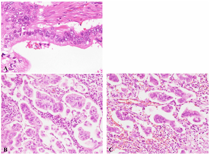
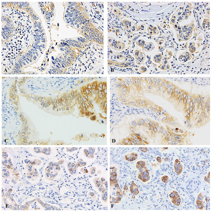
Invasive micropapillary carcinoma (IMPC) is a rare distinct histopathological subtype, characterized by the presence of carcinoma cells displaying reverse polarity. Only limited clinicopathological information is available regarding pancreatic IMPC. The aim of the present study was to clarify the clinicopathological features of pancreatic IMPC and the usefulness of protein kinase C (PKC)ζ immunostaining for the detection of reverse polarity. We reviewed 242 consecutive surgically resected specimens of pancreatic ductal adenocarcinoma and selected samples with an IMPC component. Clinicopathological characteristics were compared between the IMPC and non-IMPC groups. Immunohistochemical staining for PKCζ was performed using an autostainer. In total, 14 cases had an IMPC component (5.8%). The extent of IMPC component ranged from 5 to 20%. There were no significant differences in tumor location, T category, lymph node metastatic status, preoperative carbohydrate antigen 19-9 level, resection status and overall survival between the IMPC and non-IMPC groups. Immunostaining for PKCζ clearly showed reverse polarity of the neoplastic cells of IMPC. Although previous reports have shown that the presence of an IMPC component (>20% of the tumor) indicated poor prognosis, the present study demonstrated that presence of IMPC <20% did not suggest a worse prognosis.
浸润性微乳头状癌(IMPC)是一种罕见的独特组织病理学亚型,其特征是癌细胞呈现反向极性。关于胰腺IMPC的临床病理信息有限。本研究的目的是阐明胰腺IMPC的临床病理特征以及蛋白激酶C(PKC)ζ免疫染色对检测反向极性的实用性。我们回顾了242例连续手术切除的胰腺导管腺癌标本,并选取了具有IMPC成分的样本。比较了IMPC组和非IMPC组的临床病理特征。使用自动染色仪对PKCζ进行免疫组织化学染色。共有14例具有IMPC成分(5.8%)。IMPC成分的范围为5%至20%。IMPC组和非IMPC组在肿瘤位置、T分期、淋巴结转移状态、术前糖类抗原19-9水平、切除状态和总生存期方面无显著差异。PKCζ免疫染色清楚地显示了IMPC肿瘤细胞的反向极性。尽管先前的报告表明存在IMPC成分(>肿瘤的20%)提示预后不良,但本研究表明IMPC<20%并不提示预后更差。