• 文献检索
  • 文档翻译
  • 深度研究
  • 学术资讯
  • Suppr Zotero 插件Zotero 插件
  • 邀请有礼
  • 套餐&价格
  • 历史记录
应用&插件
Suppr Zotero 插件Zotero 插件浏览器插件Mac 客户端Windows 客户端微信小程序
定价
高级版会员购买积分包购买API积分包
服务
文献检索文档翻译深度研究API 文档MCP 服务
关于我们
关于 Suppr公司介绍联系我们用户协议隐私条款
关注我们

Suppr 超能文献

核心技术专利:CN118964589B侵权必究
粤ICP备2023148730 号-1Suppr @ 2026

文献检索

告别复杂PubMed语法,用中文像聊天一样搜索,搜遍4000万医学文献。AI智能推荐,让科研检索更轻松。

立即免费搜索

文件翻译

保留排版,准确专业,支持PDF/Word/PPT等文件格式,支持 12+语言互译。

免费翻译文档

深度研究

AI帮你快速写综述,25分钟生成高质量综述,智能提取关键信息,辅助科研写作。

立即免费体验

COVID-19 相关肝衰竭:铁过载是危险的触发因素吗?

Hepatic Failure in COVID-19: Is Iron Overload the Dangerous Trigger?

机构信息

Pathology Unit, National Institute for Infectious Diseases "Lazzaro Spallanzani"-IRCCS, 00149 Rome, Italy.

Laboratory of Electron Microscopy, National Institute for Infectious Diseases "Lazzaro Spallanzani"-IRCCS, 00149 Rome, Italy.

出版信息

Cells. 2021 May 4;10(5):1103. doi: 10.3390/cells10051103.

DOI:10.3390/cells10051103
PMID:34064487
原文链接:https://pmc.ncbi.nlm.nih.gov/articles/PMC8147922/
Abstract

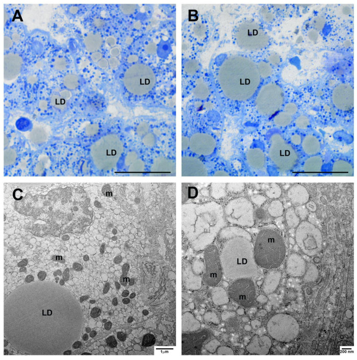
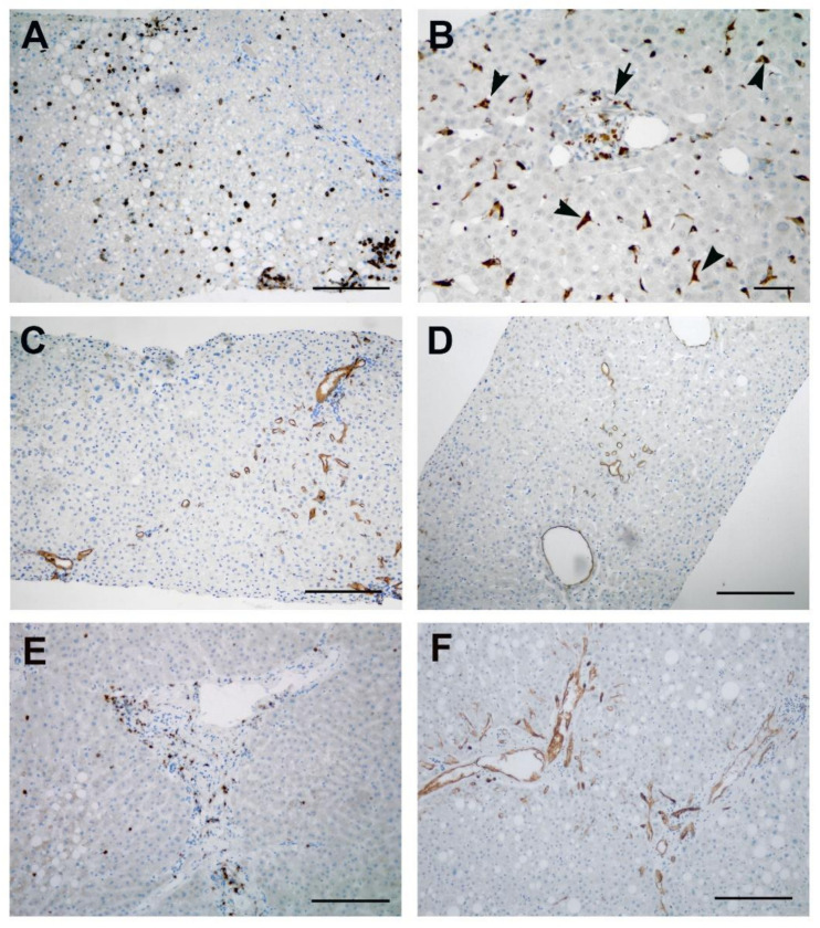
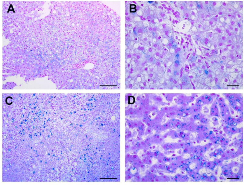
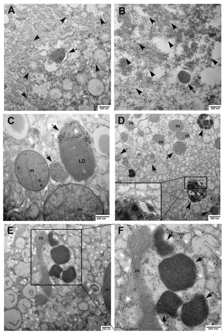
Liver injury in COVID-19 patients has progressively emerged, even in those without a history of liver disease, yet the mechanism of liver pathogenicity is still controversial. COVID-19 is frequently associated with increased serum ferritin levels, and hyperferritinemia was shown to correlate with illness severity. The liver is the major site for iron storage, and conditions of iron overload have been established to have a pathogenic role in development of liver diseases. We presented here six patients who developed severe COVID-19, with biochemical evidence of liver failure. Three cases were survived patients, who underwent liver biopsy; the other three were deceased patients, who were autopsied. None of the patients suffered underlying liver pathologies. Histopathological and ultrastructural analyses were performed. The most striking finding we demonstrated in all patients was iron accumulation into hepatocytes, associated with degenerative changes. Abundant ferritin particles were found enclosed in siderosomes, and large aggregates of hemosiderin were found, often in close contact with damaged mitochondria. Iron-caused oxidative stress may be responsible for mitochondria metabolic dysfunction. In agreement with this, association between mitochondria and lipid droplets was also found. Overall, our data suggest that hepatic iron overload could be the pathogenic trigger of liver injury associated to COVID-19.

摘要

COVID-19 患者的肝损伤逐渐显现,即使在没有肝病病史的患者中也是如此,但肝致病机制仍存在争议。COVID-19 常伴有血清铁蛋白水平升高,且高铁蛋白血症与疾病严重程度相关。肝脏是铁储存的主要部位,铁过载状态已被证实与肝脏疾病的发展具有致病作用。我们在此介绍了 6 例发生严重 COVID-19 并伴有肝功能衰竭生化证据的患者。其中 3 例为存活患者,行肝活检;另外 3 例为死亡患者,行尸检。所有患者均无潜在的肝脏疾病。进行了组织病理学和超微结构分析。我们在所有患者中最显著的发现是肝细胞内铁蓄积,伴有退行性改变。在铁蛋白小体中发现了大量的含铁血黄素,并且常与受损的线粒体紧密接触的大量含铁血黄素聚集。铁引起的氧化应激可能导致线粒体代谢功能障碍。与此一致的是,还发现了线粒体与脂滴之间的关联。总之,我们的数据表明,COVID-19 相关肝损伤的致病触发因素可能是肝内铁过载。

https://cdn.ncbi.nlm.nih.gov/pmc/blobs/4339/8147922/dc925251e16b/cells-10-01103-g005.jpg
https://cdn.ncbi.nlm.nih.gov/pmc/blobs/4339/8147922/cc5ad2d48bdb/cells-10-01103-g001.jpg
https://cdn.ncbi.nlm.nih.gov/pmc/blobs/4339/8147922/2185a7584ed2/cells-10-01103-g002.jpg
https://cdn.ncbi.nlm.nih.gov/pmc/blobs/4339/8147922/b1e2c313247e/cells-10-01103-g003.jpg
https://cdn.ncbi.nlm.nih.gov/pmc/blobs/4339/8147922/23ba816bf436/cells-10-01103-g004.jpg
https://cdn.ncbi.nlm.nih.gov/pmc/blobs/4339/8147922/dc925251e16b/cells-10-01103-g005.jpg

相似文献

1
Hepatic Failure in COVID-19: Is Iron Overload the Dangerous Trigger?COVID-19 相关肝衰竭:铁过载是危险的触发因素吗?
Cells. 2021 May 4;10(5):1103. doi: 10.3390/cells10051103.
2
Ultrastructural sequences during liver iron overload in genetic hemochromatosis.遗传性血色素沉着症肝脏铁过载时的超微结构变化序列
J Hepatol. 1997 Oct;27(4):628-38. doi: 10.1016/s0168-8278(97)80079-7.
3
Evaluating the association of serum ferritin and hepatic iron with disease severity in non-alcoholic fatty liver disease.评估血清铁蛋白和肝铁与非酒精性脂肪性肝病严重程度的相关性。
Liver Int. 2019 Jul;39(7):1325-1334. doi: 10.1111/liv.14096. Epub 2019 Mar 27.
4
Iron overload is a determinant of morbidity and mortality in adult patients with sickle cell disease.铁过载是成年镰状细胞病患者发病和死亡的一个决定因素。
Semin Hematol. 2001 Jan;38(1 Suppl 1):30-6. doi: 10.1016/s0037-1963(01)90058-7.
5
Increased Hepatic Iron Content Predicts Poor Survival in Patients With Iron Overload Who Underwent Allogeneic Hematopoietic Stem Cell Transplantation.肝铁含量增加预示接受异基因造血干细胞移植的铁过载患者生存不良。
Clin Lymphoma Myeloma Leuk. 2016 Aug;16 Suppl:S10-8. doi: 10.1016/j.clml.2016.02.005.
6
Severity of iron overload in patients with sickle cell disease receiving chronic red blood cell transfusion therapy.接受慢性红细胞输血治疗的镰状细胞病患者铁过载的严重程度。
Blood. 2000 Jul 1;96(1):76-9.
7
Iron overload in patients receiving allogeneic hematopoietic stem cell transplantation: quantification of iron burden by a superconducting quantum interference device (SQUID) and therapeutic effectiveness of phlebotomy.异体造血干细胞移植患者的铁过载:超导量子干涉仪(SQUID)定量铁负荷和放血疗法的疗效。
Biol Blood Marrow Transplant. 2010 Jan;16(1):115-22. doi: 10.1016/j.bbmt.2009.09.011. Epub 2009 Sep 18.
8
Relative contribution of iron genes, dysmetabolism and hepatitis C virus (HCV) in the pathogenesis of altered iron regulation in HCV chronic hepatitis.铁基因、代谢紊乱及丙型肝炎病毒(HCV)在HCV慢性肝炎铁调节异常发病机制中的相对作用
Haematologica. 2007 Aug;92(8):1037-42. doi: 10.3324/haematol.11281. Epub 2007 Jul 20.
9
Liver iron concentrations and urinary hepcidin in beta-thalassemia.β地中海贫血患者的肝脏铁浓度和尿铁调素
Haematologica. 2007 May;92(5):583-8. doi: 10.3324/haematol.10842.
10
Non-transferrin-bound iron and hepatic dysfunction in African dietary iron overload.非洲膳食铁过载中的非转铁蛋白结合铁与肝功能障碍
J Gastroenterol Hepatol. 1999 Feb;14(2):126-32. doi: 10.1046/j.1440-1746.1999.01830.x.

引用本文的文献

1
Mitochondria in COVID-19: from cellular and molecular perspective.新型冠状病毒肺炎中的线粒体:从细胞和分子角度看
Front Physiol. 2024 Jun 21;15:1406635. doi: 10.3389/fphys.2024.1406635. eCollection 2024.
2
Liver Iron Overload Drives COVID-19 Mortality: a Two-Sample Mendelian Randomization Study.肝脏铁过载导致 COVID-19 死亡率升高:两样本 Mendelian 随机研究。
Biol Trace Elem Res. 2024 Jun;202(6):2509-2517. doi: 10.1007/s12011-023-03878-8. Epub 2023 Oct 9.
3
COVID-19 and β-thalassemia: in lieu of evidence and vague nexus.COVID-19 和 β-地中海贫血:缺乏证据和模糊的联系。

本文引用的文献

1
Evidences for lipid involvement in SARS-CoV-2 cytopathogenesis.脂质在 SARS-CoV-2 细胞病变发生中的作用证据。
Cell Death Dis. 2021 Mar 12;12(3):263. doi: 10.1038/s41419-021-03527-9.
2
COVID-19-associated liver injury: from bedside to bench.COVID-19 相关肝损伤:从床边到实验室。
J Gastroenterol. 2021 Mar;56(3):218-230. doi: 10.1007/s00535-021-01760-9. Epub 2021 Feb 1.
3
Multi-organ proteomic landscape of COVID-19 autopsies.COVID-19 尸检的多器官蛋白质组学图谱。
Ann Hematol. 2024 May;103(5):1423-1433. doi: 10.1007/s00277-023-05346-8. Epub 2023 Jul 5.
4
COVID-19 induced liver injury from a new perspective: Mitochondria.从新视角看 COVID-19 诱导的肝损伤:线粒体。
Mitochondrion. 2023 May;70:103-110. doi: 10.1016/j.mito.2023.04.001. Epub 2023 Apr 11.
5
Autopsies Revealed Pathological Features of COVID-19 in Unvaccinated vs. Vaccinated Patients.尸检揭示了未接种疫苗与接种疫苗的新冠患者的病理特征。
Biomedicines. 2023 Feb 14;11(2):551. doi: 10.3390/biomedicines11020551.
6
Liver pathology in COVID-19 related death and leading role of autopsy in the pandemic.COVID-19 相关死亡的肝脏病理学和尸检在大流行中的主导作用。
World J Gastroenterol. 2023 Jan 7;29(1):200-220. doi: 10.3748/wjg.v29.i1.200.
7
Digestive system infection by SARS‑CoV‑2: Entry mechanism, clinical symptoms and expression of major receptors (Review).消化系统感染 SARS-CoV-2:进入机制、临床症状和主要受体的表达(综述)。
Int J Mol Med. 2023 Mar;51(3). doi: 10.3892/ijmm.2023.5222. Epub 2023 Jan 20.
8
Liver injury in COVID-19: A minireview.新型冠状病毒肺炎相关肝损伤:综述
World J Gastroenterol. 2022 Dec 21;28(47):6716-6731. doi: 10.3748/wjg.v28.i47.6716.
9
Understanding Long COVID; Mitochondrial Health and Adaptation-Old Pathways, New Problems.了解新冠后长期症状;线粒体健康与适应——古老的途径,新的问题。
Biomedicines. 2022 Dec 2;10(12):3113. doi: 10.3390/biomedicines10123113.
10
Metabolic Reprogramming in SARS-CoV-2 Infection Impacts the Outcome of COVID-19 Patients.SARS-CoV-2 感染中的代谢重编程影响 COVID-19 患者的结局。
Front Immunol. 2022 Jul 11;13:936106. doi: 10.3389/fimmu.2022.936106. eCollection 2022.
Cell. 2021 Feb 4;184(3):775-791.e14. doi: 10.1016/j.cell.2021.01.004. Epub 2021 Jan 9.
4
Ferroptosis: mechanisms, biology and role in disease.铁死亡:机制、生物学及其在疾病中的作用
Nat Rev Mol Cell Biol. 2021 Apr;22(4):266-282. doi: 10.1038/s41580-020-00324-8. Epub 2021 Jan 25.
5
Persisting alterations of iron homeostasis in COVID-19 are associated with non-resolving lung pathologies and poor patients' performance: a prospective observational cohort study.在 COVID-19 中持续存在的铁稳态紊乱与肺部病理无法消退和患者预后不良有关:一项前瞻性观察队列研究。
Respir Res. 2020 Oct 21;21(1):276. doi: 10.1186/s12931-020-01546-2.
6
Clinical characteristics and risk factors of liver injury in COVID-19: a retrospective cohort study from Wuhan, China.新型冠状病毒肺炎患者肝损伤的临床特征及危险因素:一项来自中国武汉的回顾性队列研究。
Hepatol Int. 2020 Sep;14(5):723-732. doi: 10.1007/s12072-020-10075-5. Epub 2020 Oct 7.
7
Postmortem Findings in Italian Patients With COVID-19: A Descriptive Full Autopsy Study of Cases With and Without Comorbidities.意大利 COVID-19 患者的尸检结果:有和无合并症病例的描述性全面尸检研究。
J Infect Dis. 2020 Nov 9;222(11):1807-1815. doi: 10.1093/infdis/jiaa578.
8
Taxifolin ameliorates iron overload-induced hepatocellular injury: Modulating PI3K/AKT and p38 MAPK signaling, inflammatory response, and hepatocellular regeneration.圣约翰草提取物可改善铁过载诱导的肝细胞损伤:调控 PI3K/AKT 和 p38 MAPK 信号通路、炎症反应和肝细胞再生。
Chem Biol Interact. 2020 Oct 1;330:109230. doi: 10.1016/j.cbi.2020.109230. Epub 2020 Aug 20.
9
Hepatic pathology in patients dying of COVID-19: a series of 40 cases including clinical, histologic, and virologic data.COVID-19 死亡患者的肝脏病理学:包含临床、组织学和病毒学数据的 40 例系列病例。
Mod Pathol. 2020 Nov;33(11):2147-2155. doi: 10.1038/s41379-020-00649-x. Epub 2020 Aug 13.
10
Prevalence and Predictive Value of Anemia and Dysregulated Iron Homeostasis in Patients with COVID-19 Infection.新型冠状病毒肺炎感染患者贫血和铁稳态失调的患病率及预测价值
J Clin Med. 2020 Jul 29;9(8):2429. doi: 10.3390/jcm9082429.