CNR Neuroscience Institute, 56124 Pisa, Italy.
Department of Biology, University of Pisa, 56126 Pisa, Italy.
Int J Mol Sci. 2021 May 20;22(10):5381. doi: 10.3390/ijms22105381.
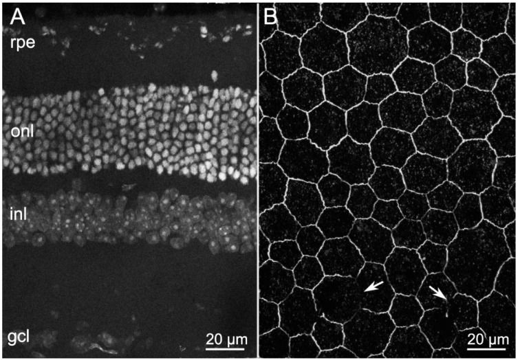
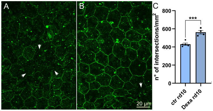
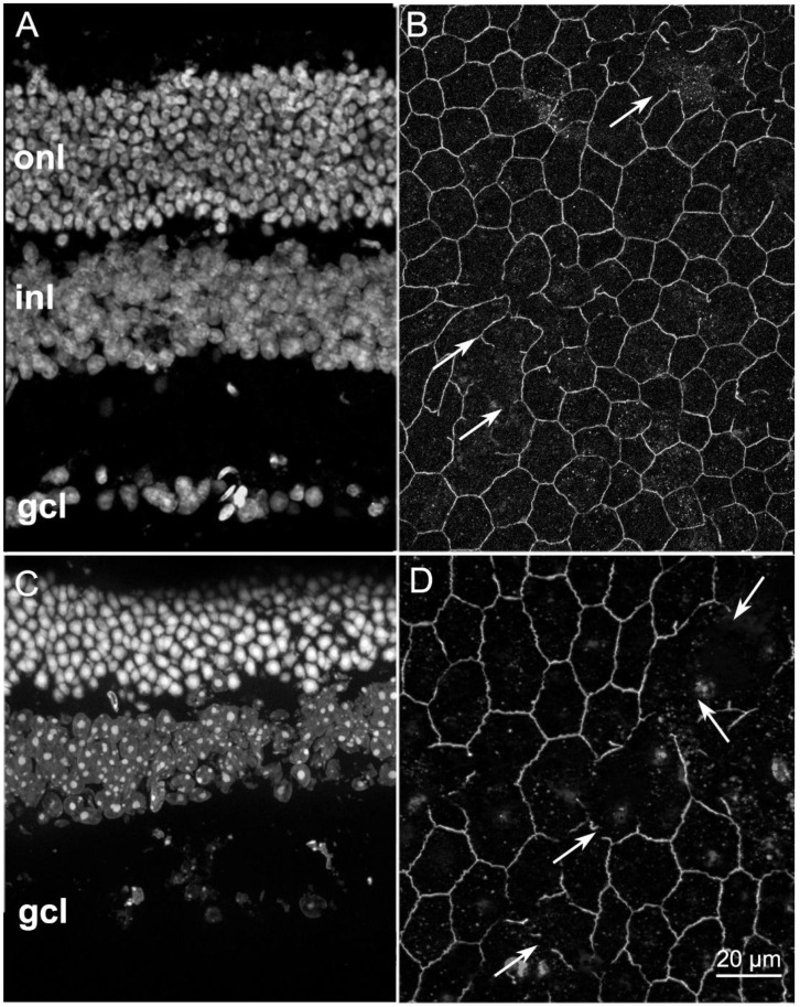
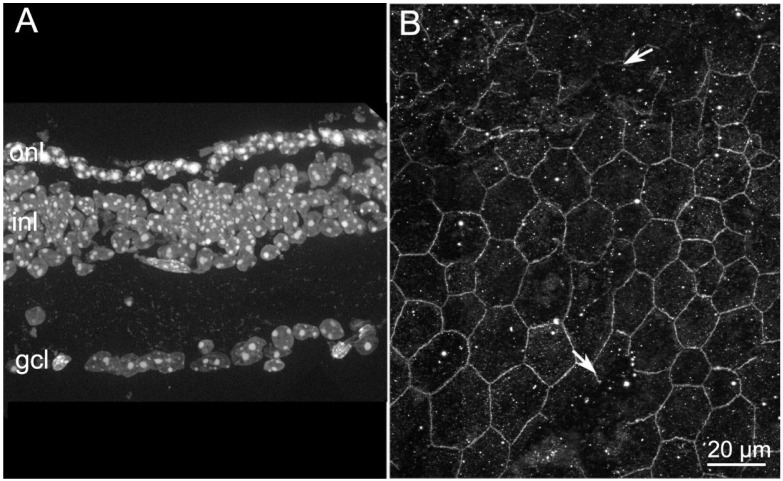
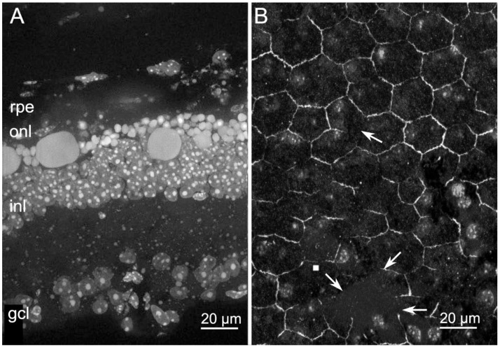
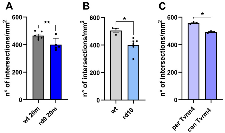
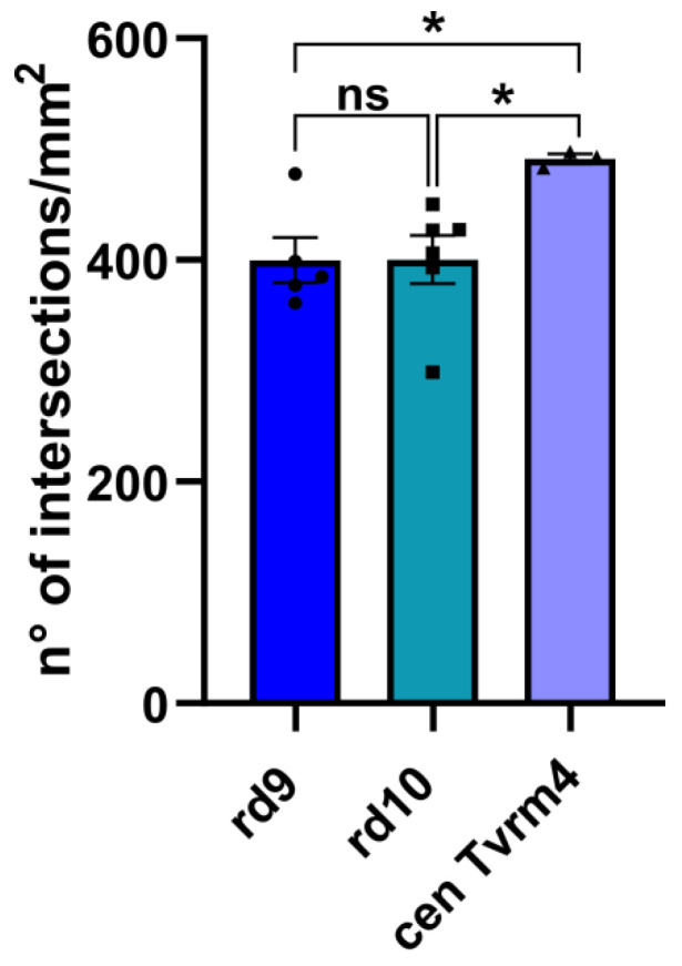
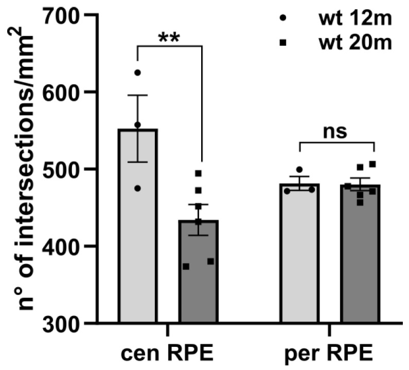
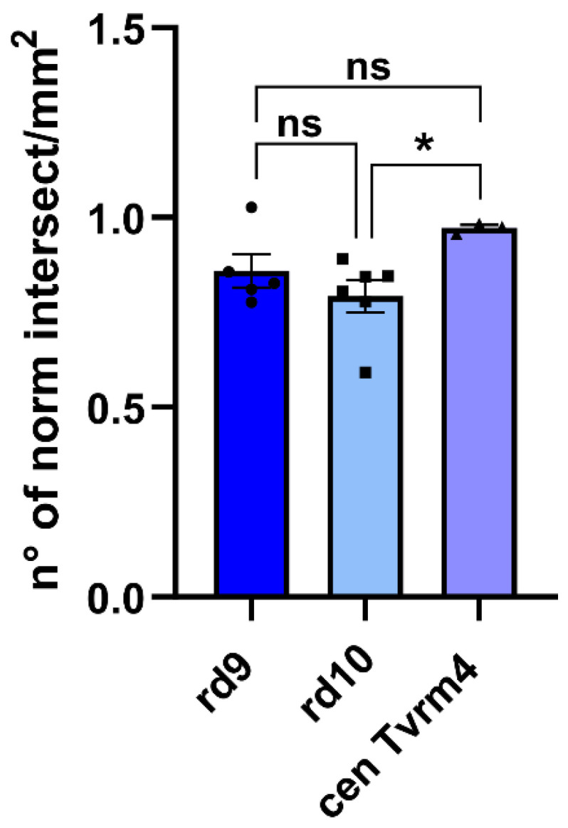
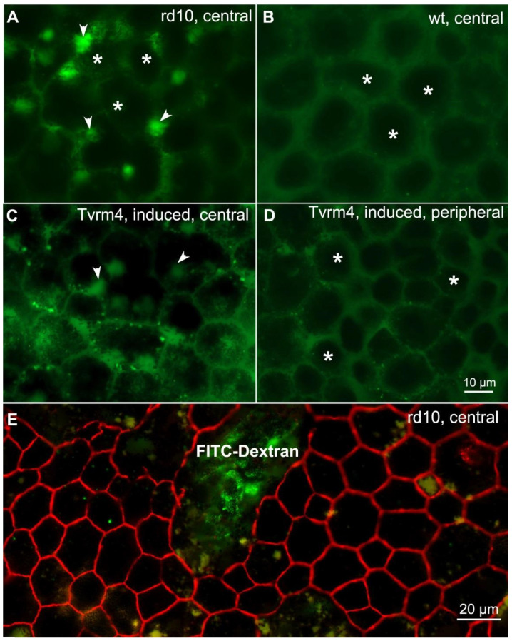
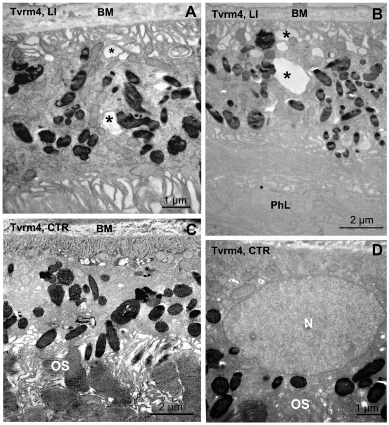
In retinitis pigmentosa (RP), one of many possible genetic mutations causes rod degeneration, followed by cone secondary death leading to blindness. Accumulating evidence indicates that rod death triggers multiple, non-cell-autonomous processes, which include oxidative stress and inflammation/immune responses, all contributing to cone demise. Inflammation relies on local microglia and recruitment of immune cells, reaching the retina through breakdowns of the inner blood retinal barrier (iBRB). Leakage in the inner retina vasculature suggests similarly altered outer BRB, formed by junctions between retinal pigment epithelium (RPE) cells, which are crucial for retinal homeostasis, immune response, and privilege. We investigated the RPE structural integrity in three models of RP (rd9, rd10, and Tvrm4 mice) by immunostaining for zonula occludens-1 (ZO-1), an essential regulatory component of tight junctions. Quantitative image analysis demonstrated discontinuities in ZO-1 profiles in all mutants, despite different degrees of photoreceptor loss. ZO-1 interruption zones corresponded to leakage of in vivo administered, fluorescent dextran through the choroid-RPE interface, demonstrating barrier dysfunction. Dexamethasone, administered to rd10 mice for rescuing cones, also rescued RPE structure. Thus, previously undetected, stereotyped abnormalities occur in the RPE of RP mice; pharmacological targeting of inflammation supports a feedback loop leading to simultaneous protection of cones and the RPE.
在视网膜色素变性(RP)中,许多可能的基因突变之一导致视杆细胞退化,随后视锥细胞继发性死亡导致失明。越来越多的证据表明,视杆细胞死亡引发了多种非细胞自主过程,包括氧化应激和炎症/免疫反应,所有这些都导致视锥细胞死亡。炎症依赖于局部小胶质细胞和免疫细胞的募集,通过内血视网膜屏障(iBRB)的破裂到达视网膜。内视网膜血管的渗漏表明类似的外 BRB 也发生了改变,外 BRB 由视网膜色素上皮(RPE)细胞之间的连接形成,对于视网膜内稳态、免疫反应和特权至关重要。我们通过免疫染色检测紧密连接的重要调节成分封闭蛋白-1(ZO-1),研究了三种 RP 模型(rd9、rd10 和 Tvrm4 小鼠)中的 RPE 结构完整性。定量图像分析表明,尽管感光细胞丧失程度不同,但所有突变体的 ZO-1 图谱都存在不连续性。ZO-1 中断区与体内给予的荧光葡聚糖通过脉络膜-RPE 界面渗漏相对应,表明屏障功能障碍。对 rd10 小鼠给予地塞米松以挽救视锥细胞,也挽救了 RPE 结构。因此,在 RP 小鼠的 RPE 中出现了以前未检测到的刻板异常;炎症的药物靶向支持导致同时保护视锥细胞和 RPE 的反馈回路。