• 文献检索
  • 文档翻译
  • 深度研究
  • 学术资讯
  • Suppr Zotero 插件Zotero 插件
  • 邀请有礼
  • 套餐&价格
  • 历史记录
应用&插件
Suppr Zotero 插件Zotero 插件浏览器插件Mac 客户端Windows 客户端微信小程序
定价
高级版会员购买积分包购买API积分包
服务
文献检索文档翻译深度研究API 文档MCP 服务
关于我们
关于 Suppr公司介绍联系我们用户协议隐私条款
关注我们

Suppr 超能文献

核心技术专利:CN118964589B侵权必究
粤ICP备2023148730 号-1Suppr @ 2026

文献检索

告别复杂PubMed语法,用中文像聊天一样搜索,搜遍4000万医学文献。AI智能推荐,让科研检索更轻松。

立即免费搜索

文件翻译

保留排版,准确专业,支持PDF/Word/PPT等文件格式,支持 12+语言互译。

免费翻译文档

深度研究

AI帮你快速写综述,25分钟生成高质量综述,智能提取关键信息,辅助科研写作。

立即免费体验

CT/MRI版LI-RADS v2018与CEUS版LI-RADS v2017——能否整合?

CT/MRI LI-RADS v2018 vs. CEUS LI-RADS v2017-Can Things Be Put Together?

作者信息

Caraiani Cosmin, Boca Bianca, Bura Vlad, Sparchez Zeno, Dong Yi, Dietrich Christoph

机构信息

Department of Medical Imaging, "Iuliu Hațieganu" University of Medicine and Pharmacy Cluj-Napoca, 400012 Cluj-Napoca, Romania.

Department of Radiology, County Clinical Emergency Hospital Cluj-Napoca, 400006 Cluj-Napoca, Romania.

出版信息

Biology (Basel). 2021 May 6;10(5):412. doi: 10.3390/biology10050412.

DOI:10.3390/biology10050412
PMID:34066607
原文链接:https://pmc.ncbi.nlm.nih.gov/articles/PMC8148521/
Abstract

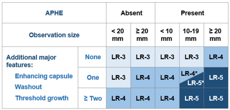
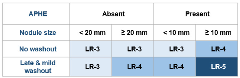
Different LI-RADS core documents were released for CEUS and for CT/MRI. Both documents rely on major and ancillary diagnostic criteria. The present paper offers an exhaustive comparison of the two documents focusing on the similarities, but especially on the differences, complementarity, and added value of imaging techniques in classifying liver nodules in cirrhotic livers. The major diagnostic criteria are defined, and the sensitivity and specificity of each major diagnostic criteria are presented according to the literature. The existing differences between techniques in assessing the major diagnostic features can be then exploited in order to ensure a better classification and a better clinical management of liver nodules in cirrhotic livers. Ancillary features depend on the imaging technique used, and their presence can upgrade or downgrade the LI-RADS score of an observation, but only as far as LI-RADS 4. MRI is the imaging technique that provides the greatest number of ancillary features, whereas CEUS has fewer ancillary features than other imaging techniques. In the final part of the manuscript, some recommendations are made by the authors in order to guidephysicians as to when adding another imaging technique can be helpful in managing liver nodules in cirrhotic livers.

摘要

针对超声造影(CEUS)以及CT/MRI发布了不同的肝脏影像报告和数据系统(LI-RADS)核心文档。这两份文档均依赖主要和辅助诊断标准。本文对两份文档进行了详尽比较,重点关注它们的相似之处,但尤其关注在对肝硬化肝脏中的肝结节进行分类时,成像技术的差异、互补性及附加价值。明确了主要诊断标准,并根据文献列出了各主要诊断标准的敏感性和特异性。评估主要诊断特征时技术上的现有差异随后可被利用,以确保对肝硬化肝脏中的肝结节进行更好的分类和临床管理。辅助特征取决于所使用的成像技术,其存在可提高或降低某个观察结果的LI-RADS评分,但仅限于LI-RADS 4类。MRI是提供辅助特征数量最多的成像技术,而CEUS的辅助特征比其他成像技术少。在本文的最后部分,作者给出了一些建议,以指导医生在何时增加另一种成像技术有助于管理肝硬化肝脏中的肝结节。

https://cdn.ncbi.nlm.nih.gov/pmc/blobs/e8a3/8148521/0b2e84f5b607/biology-10-00412-g006.jpg
https://cdn.ncbi.nlm.nih.gov/pmc/blobs/e8a3/8148521/9c35e9ae6149/biology-10-00412-g001.jpg
https://cdn.ncbi.nlm.nih.gov/pmc/blobs/e8a3/8148521/64a83121c0d7/biology-10-00412-g002.jpg
https://cdn.ncbi.nlm.nih.gov/pmc/blobs/e8a3/8148521/9e6df6cf9e24/biology-10-00412-g003.jpg
https://cdn.ncbi.nlm.nih.gov/pmc/blobs/e8a3/8148521/1b03560a4877/biology-10-00412-g004.jpg
https://cdn.ncbi.nlm.nih.gov/pmc/blobs/e8a3/8148521/88804a423239/biology-10-00412-g005.jpg
https://cdn.ncbi.nlm.nih.gov/pmc/blobs/e8a3/8148521/0b2e84f5b607/biology-10-00412-g006.jpg

相似文献

1
CT/MRI LI-RADS v2018 vs. CEUS LI-RADS v2017-Can Things Be Put Together?CT/MRI版LI-RADS v2018与CEUS版LI-RADS v2017——能否整合?
Biology (Basel). 2021 May 6;10(5):412. doi: 10.3390/biology10050412.
2
Diagnostic performance of MR for hepatocellular carcinoma based on LI-RADS v2018, compared with v2017.基于 LI-RADS v2018 的肝细胞癌的 MRI 诊断性能,与 v2017 相比。
J Magn Reson Imaging. 2019 Sep;50(3):746-755. doi: 10.1002/jmri.26640. Epub 2019 Jan 15.
3
Comparison of the diagnostic performance of the 2017 and 2018 versions of LI-RADS for hepatocellular carcinoma on gadoxetic acid enhanced MRI.对比钆塞酸增强 MRI 上 2017 版和 2018 版 LI-RADS 对肝细胞癌的诊断性能。
Clin Radiol. 2020 Apr;75(4):319.e1-319.e9. doi: 10.1016/j.crad.2019.11.004. Epub 2019 Dec 16.
4
Critical analysis of major and ancillary features of LI-RADS v2018 in the differentiation of small (≤ 2 cm) hepatocellular carcinoma from dysplastic nodules with gadobenate dimeglumine-enhanced magnetic resonance imaging.LI-RADS v2018 主要特征和辅助特征的批判性分析在使用钆贝葡胺增强磁共振成像鉴别≤2cm 的小肝癌与异型增生结节中的作用。
Eur Rev Med Pharmacol Sci. 2019 Sep;23(18):7786-7801. doi: 10.26355/eurrev_201909_18988.
5
LI-RADS Version 2017 versus Version 2018: Diagnosis of Hepatocellular Carcinoma on Gadoxetate Disodium-enhanced MRI.LI-RADS 版本 2017 与版本 2018:钆塞酸二钠增强 MRI 诊断肝细胞癌。
Radiology. 2019 Sep;292(3):655-663. doi: 10.1148/radiol.2019182867. Epub 2019 Jul 16.
6
Contrast-enhanced ultrasound LI-RADS 2017: comparison with CT/MRI LI-RADS.对比增强超声 LI-RADS 2017:与 CT/MRI LI-RADS 的比较。
Eur Radiol. 2021 Feb;31(2):847-854. doi: 10.1007/s00330-020-07159-z. Epub 2020 Aug 15.
7
Diagnostic Accuracy of CEUS LI-RADS for the Characterization of Liver Nodules 20 mm or Smaller in Patients at Risk for Hepatocellular Carcinoma.CEUS LI-RADS 在肝癌风险患者中对 20mm 或更小的肝脏结节的特征诊断准确性。
Radiology. 2020 Feb;294(2):329-339. doi: 10.1148/radiol.2019191086. Epub 2019 Dec 3.
8
Liver Imaging Reporting and Data System Version 2018: Impact on Categorization and Hepatocellular Carcinoma Staging.肝脏影像报告和数据系统 2018 年版:对分类和肝细胞癌分期的影响。
Liver Transpl. 2019 Oct;25(10):1488-1502. doi: 10.1002/lt.25614. Epub 2019 Sep 9.
9
Diagnostic Performance of LI-RADS Version 2018, LI-RADS Version 2017, and OPTN Criteria for Hepatocellular Carcinoma.LI-RADS 版本 2018、LI-RADS 版本 2017 和 OPTN 标准用于诊断肝细胞癌的性能比较。
AJR Am J Roentgenol. 2020 Nov;215(5):1085-1092. doi: 10.2214/AJR.20.22772. Epub 2020 Sep 2.
10
White paper of the Society of Abdominal Radiology hepatocellular carcinoma diagnosis disease-focused panel on LI-RADS v2018 for CT and MRI.美国腹部放射学会肝癌诊断疾病聚焦专家组关于 CT 和 MRI 的 LI-RADS v2018 白皮书。
Abdom Radiol (NY). 2018 Oct;43(10):2625-2642. doi: 10.1007/s00261-018-1744-4.

引用本文的文献

1
The role of contrast-enhanced ultrasound in the radiological classification of liver observations identified by CT and MRI.超声造影在CT和MRI所发现肝脏病变的影像学分类中的作用。
Radiol Med. 2025 Mar 24. doi: 10.1007/s11547-025-01995-z.
2
Contrast-enhanced US of High-Risk Indeterminate Focal Liver Observations Categorized as LR-4 or LR-M at CT/MRI.CT/MRI上分类为LR-4或LR-M的高风险不确定局灶性肝脏病变的超声造影检查
Radiology. 2025 Jan;314(1):e240916. doi: 10.1148/radiol.240916.
3
Gadoxetic acid disodium (Gd-EOB-DTPA) contrast-enhanced abbreviated magnetic resonance imaging (MRI) for hepatocellular carcinoma surveillance in at-risk patients: a multi-center study in China.

本文引用的文献

1
Comparison of Contrast-Enhanced Ultrasound versus Contrast-Enhanced Magnetic Resonance Imaging for the Diagnosis of Focal Liver Lesions Using the Liver Imaging Reporting and Data System.对比增强超声与对比增强磁共振成像在肝脏影像报告与数据系统诊断局灶性肝脏病变中的比较。
Ultrasound Med Biol. 2020 May;46(5):1216-1223. doi: 10.1016/j.ultrasmedbio.2020.01.023. Epub 2020 Feb 27.
2
Resolution of indeterminate MRI with CEUS in patients at high risk for hepatocellular carcinoma.CEUS 对高度疑似肝细胞癌患者 MRI 不确定结果的诊断价值。
Abdom Radiol (NY). 2020 Jan;45(1):123-133. doi: 10.1007/s00261-019-02181-2.
3
钆塞酸二钠(Gd-EOB-DTPA)增强简化磁共振成像(MRI)用于高危患者肝细胞癌监测:一项中国多中心研究
Quant Imaging Med Surg. 2024 Dec 5;14(12):8520-8537. doi: 10.21037/qims-24-941. Epub 2024 Oct 24.
4
Unraveling distinctions between contrast-enhanced ultrasound and CT/MRI for liver mass diagnosis.解析超声造影与CT/MRI在肝脏肿块诊断中的差异
Ultrasonography. 2025 Jan;44(1):19-30. doi: 10.14366/usg.24163. Epub 2024 Oct 16.
5
Combining CEUS and CT/MRI LI-RADS major imaging features: diagnostic accuracy for classification of indeterminate liver observations in patients at risk for HCC.结合CEUS与CT/MRI LI-RADS主要影像学特征:对肝癌高危患者肝脏不确定病变分类的诊断准确性
Abdom Radiol (NY). 2025 May;50(5):2066-2077. doi: 10.1007/s00261-024-04625-w. Epub 2024 Oct 23.
6
Dynamic contrast-enhanced ultrasound findings of focal nodular hyperplasia-like nodules in cirrhosis: a description of two cases and literature analysis.肝硬化中局灶性结节样增生样结节的动态对比增强超声表现:两例病例描述及文献分析
Quant Imaging Med Surg. 2024 Feb 1;14(2):2089-2095. doi: 10.21037/qims-23-907. Epub 2024 Jan 9.
7
Compared with CT/MRI LI-RADS, whether CEUS LI-RADS is worth popularizing in diagnosis of hepatocellular carcinoma?-a direct head-to-head meta-analysis.与CT/MRI肝脏影像报告和数据系统(LI-RADS)相比,对比增强超声(CEUS)肝脏影像报告和数据系统在肝细胞癌诊断中是否值得推广?——一项直接的头对头荟萃分析。
Quant Imaging Med Surg. 2023 Aug 1;13(8):4919-4932. doi: 10.21037/qims-22-1383. Epub 2023 May 24.
8
Overlap syndrome of autoimmune hepatitis and primary biliary cholangitis complicated with atypical hepatocellular carcinoma: a case report.自身免疫性肝炎和原发性胆汁性胆管炎重叠综合征合并非典型肝细胞癌:一例报告。
J Med Case Rep. 2023 Jul 25;17(1):328. doi: 10.1186/s13256-023-03932-y.
9
Incidental Findings in Pediatric Patients: How to Manage Liver Incidentaloma in Pediatric Patients.儿科患者的偶然发现:如何处理儿科患者的肝脏偶然瘤
Cancers (Basel). 2023 Apr 18;15(8):2360. doi: 10.3390/cancers15082360.
10
Spontaneously Ruptured Hepatocellular Carcinoma: Computed Tomography-Based Assessment.自发性破裂肝细胞癌:基于计算机断层扫描的评估
Diagnostics (Basel). 2023 Mar 7;13(6):1021. doi: 10.3390/diagnostics13061021.
Additional value of contrast-enhanced ultrasound (CEUS) on arterial phase non-hyperenhancement observations (≥ 2 cm) of CT/MRI for high-risk patients: focusing on the CT/MRI LI-RADS categories LR-3 and LR-4.
增强超声(CEUS)对 CT/MRI 动脉期非高增强观察(≥2cm)的附加价值(高危患者):重点关注 CT/MRI LI-RADS 类别 LR-3 和 LR-4。
Abdom Radiol (NY). 2020 Jan;45(1):55-63. doi: 10.1007/s00261-019-02132-x.
4
LI-RADS: a conceptual and historical review from its beginning to its recent integration into AASLD clinical practice guidance.肝脏影像报告和数据系统(LI-RADS):从其起源到最近纳入美国肝病研究学会(AASLD)临床实践指南的概念与历史回顾
J Hepatocell Carcinoma. 2019 Feb 5;6:49-69. doi: 10.2147/JHC.S186239. eCollection 2019.
5
LI-RADS Version 2018 Ancillary Features at MRI.LI-RADS Version 2018 磁共振成像辅助特征。
Radiographics. 2018 Nov-Dec;38(7):1973-2001. doi: 10.1148/rg.2018180052. Epub 2018 Oct 5.
6
LI-RADS for CT diagnosis of hepatocellular carcinoma: performance of major and ancillary features.LI-RADS 用于 CT 诊断肝细胞癌:主要特征和辅助特征的性能。
Abdom Radiol (NY). 2019 Feb;44(2):517-528. doi: 10.1007/s00261-018-1762-2.
7
LI-RADS version 2018: What is new and what does this mean to my radiology reports?《肝脏影像报告和数据系统(LI-RADS)2018版:有哪些新内容以及这对我的放射学报告意味着什么?》
Abdom Radiol (NY). 2019 Jan;44(1):41-42. doi: 10.1007/s00261-018-1730-x.
8
LI-RADS for MR Imaging Diagnosis of Hepatocellular Carcinoma: Performance of Major and Ancillary Features.LI-RADS 用于肝细胞癌的 MRI 诊断:主要特征和次要特征的性能。
Radiology. 2018 Jul;288(1):118-128. doi: 10.1148/radiol.2018171678. Epub 2018 Apr 10.
9
Surveillance Imaging and Alpha Fetoprotein for Early Detection of Hepatocellular Carcinoma in Patients With Cirrhosis: A Meta-analysis.肝硬化患者肝细胞癌早期检测的监测成像和甲胎蛋白:一项荟萃分析。
Gastroenterology. 2018 May;154(6):1706-1718.e1. doi: 10.1053/j.gastro.2018.01.064. Epub 2018 Feb 6.
10
Evidence Supporting LI-RADS Major Features for CT- and MR Imaging-based Diagnosis of Hepatocellular Carcinoma: A Systematic Review.支持LI-RADS主要特征用于基于CT和MRI成像诊断肝细胞癌的证据:一项系统评价
Radiology. 2018 Jan;286(1):29-48. doi: 10.1148/radiol.2017170554. Epub 2017 Nov 21.