Looprasertkul Sasikarn, Sereemaspun Amornpun, Kitkumthorn Nakarin, Sooklert Kanidta, Sarachana Tewarit, Jindatip Depicha
Department of Anatomy, Faculty of Medicine, Chulalongkorn University, 1873 Rama 4 Rd., Wangmai, Pathumwan, Bangkok 10330, Thailand.
Nanomedicine Research Unit, Department of Anatomy, Faculty of Medicine, Chulalongkorn University, Bangkok 10330, Thailand.
Pharmaceutics. 2021 May 17;13(5):738. doi: 10.3390/pharmaceutics13050738.
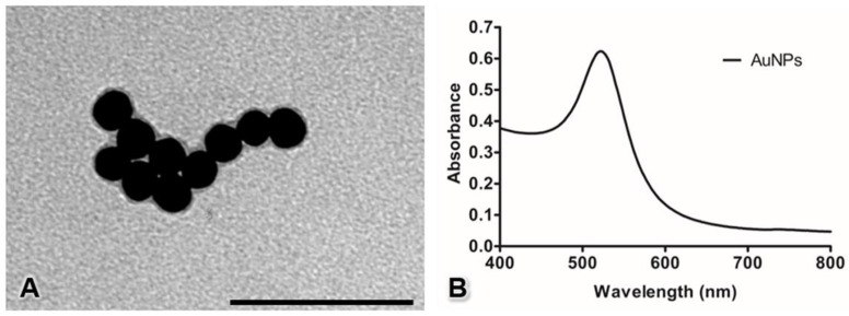
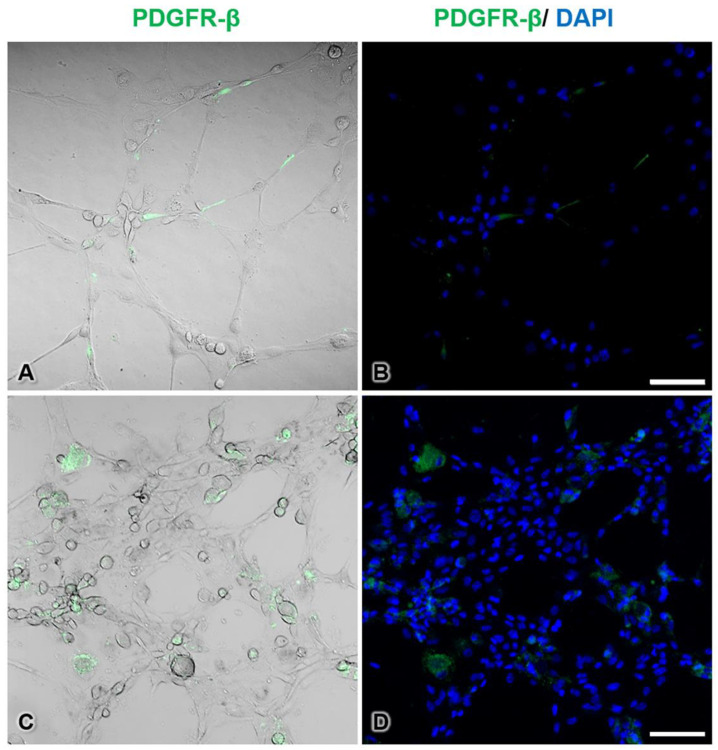
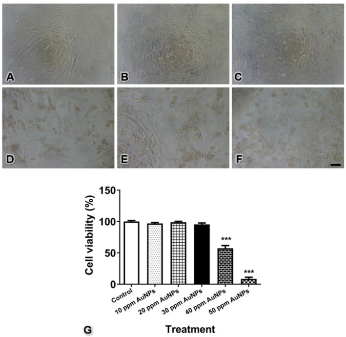
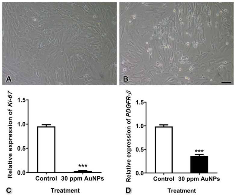
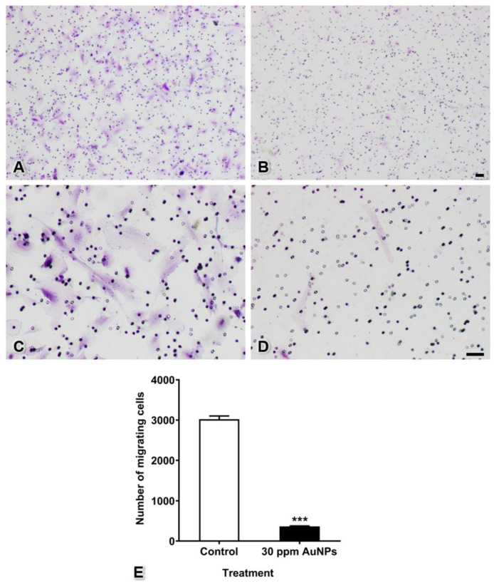
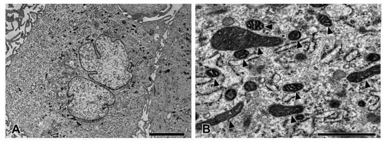
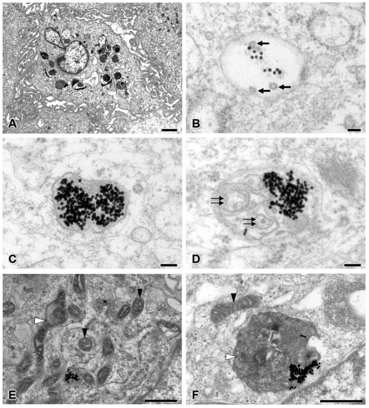
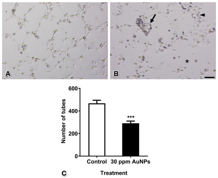
Gold nanoparticles (AuNPs) are used for diagnostic and therapeutic purposes, especially antiangiogenesis, which are accomplished via inhibition of endothelial cell proliferation, migration, and tube formation. However, no research has been performed on the effects of AuNPs in pericytes, which play vital roles in endothelial cell functions and capillary tube formation during physiological and pathological processes. Therefore, the effects of AuNPs on the morphology and functions of pericytes need to be elucidated. This study treated human placental pericytes in monoculture with 20 nm AuNPs at a concentration of 30 ppm. Ki-67 and platelet-derived growth factor receptor-β (PDGFR-β) mRNA expression was measured using real-time reverse transcription-quantitative polymerase chain reaction (RT-qPCR). Cell migration was assessed by Transwell migration assay. The fine structures of pericytes were observed by transmission electron microscopy. In addition, 30 ppm AuNP-treated pericytes and intact human umbilical vein endothelial cells were cocultured on Matrigel to form three-dimensional (3D) capillary tubes. The results demonstrated that AuNPs significantly inhibited proliferation, reduced PDGFR-β mRNA expression, and decreased migration in pericytes. Ultrastructural analysis of pericytes revealed AuNPs in late endosomes, autolysosomes, and mitochondria. Remarkably, many mitochondria were swollen or damaged. Additionally, capillary tube formation was reduced. We found that numerous pericytes on 3D capillary tubes were round and did not extend their processes along the tubes, which resulted in more incomplete tube formation in the treatment group compared with the control group. In summary, AuNPs can affect pericyte proliferation, PDGFR-β mRNA expression, migration, morphology, and capillary tube formation. The findings highlight the possible application of AuNPs in pericyte-targeted therapy for antiangiogenesis.
金纳米颗粒(AuNPs)被用于诊断和治疗目的,尤其是抗血管生成,这是通过抑制内皮细胞增殖、迁移和管腔形成来实现的。然而,尚未有关于AuNPs对周细胞影响的研究,而周细胞在生理和病理过程中的内皮细胞功能及毛细血管管腔形成中起着至关重要的作用。因此,需要阐明AuNPs对周细胞形态和功能的影响。本研究用浓度为30 ppm的20 nm AuNPs处理单培养的人胎盘周细胞。使用实时逆转录定量聚合酶链反应(RT-qPCR)测量Ki-67和血小板衍生生长因子受体-β(PDGFR-β)mRNA表达。通过Transwell迁移试验评估细胞迁移。通过透射电子显微镜观察周细胞的精细结构。此外,将经30 ppm AuNP处理的周细胞和完整的人脐静脉内皮细胞在基质胶上共培养以形成三维(3D)毛细血管管腔。结果表明,AuNPs显著抑制周细胞增殖,降低PDGFR-β mRNA表达,并减少其迁移。周细胞的超微结构分析显示晚期内体、自噬溶酶体和线粒体中有AuNPs。值得注意的是,许多线粒体肿胀或受损。此外,毛细血管管腔形成减少。我们发现3D毛细血管管腔上的许多周细胞呈圆形,且其突起未沿管腔延伸,这导致与对照组相比,治疗组的管腔形成更不完整。总之,AuNPs可影响周细胞增殖、PDGFR-β mRNA表达、迁移、形态及毛细血管管腔形成。这些发现突出了AuNPs在周细胞靶向抗血管生成治疗中的可能应用。