Foti Pietro Valerio, Travali Mario, Farina Renato, Palmucci Stefano, Spatola Corrado, Liardo Rocco Luca Emanuele, Milazzotto Roberto, Raffaele Luigi, Salamone Vincenzo, Caltabiano Rosario, Broggi Giuseppe, Puzzo Lidia, Russo Andrea, Reibaldi Michele, Longo Antonio, Vigneri Paolo, Avitabile Teresio, Ettorre Giovani Carlo, Basile Antonio
Department of Medical Surgical Sciences and Advanced Technologies "G.F. Ingrassia" - Radiology I Unit, University Hospital Policlinico "G. Rodolico-San Marco", Via Santa Sofia, 78 - 95123, Catania, Italy.
Department of Medical Surgical Sciences and Advanced Technologies "G.F. Ingrassia" - Section of Anatomic Pathology, University of Catania, Via Santa Sofia, 78 - 95123, Catania, Italy.
Insights Imaging. 2021 Jun 4;12(1):67. doi: 10.1186/s13244-021-01001-w.
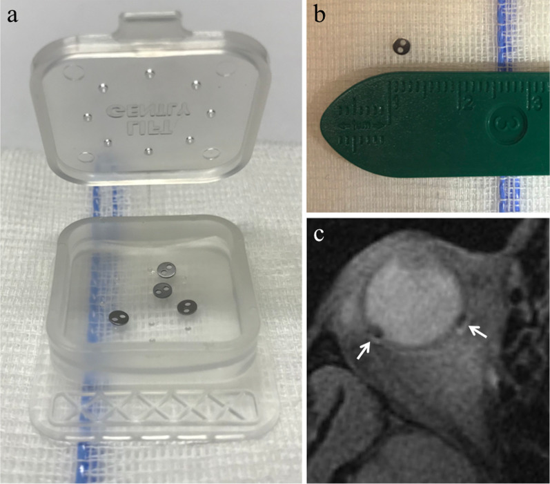
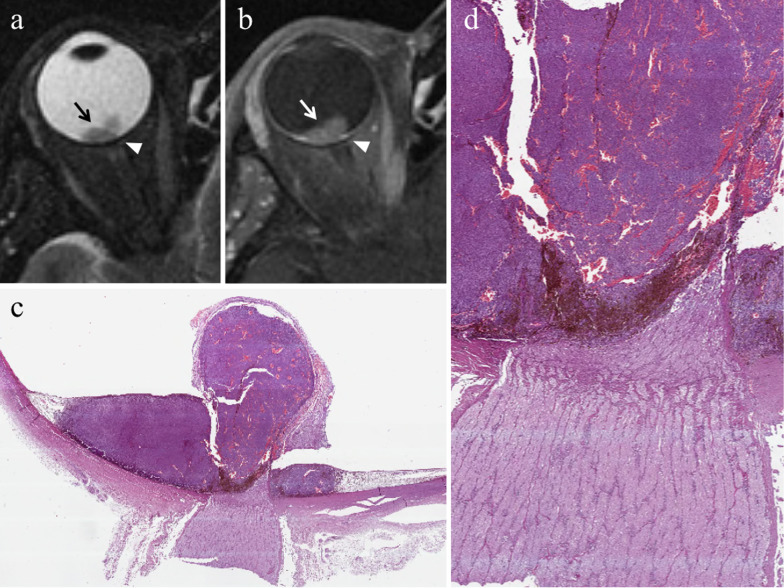
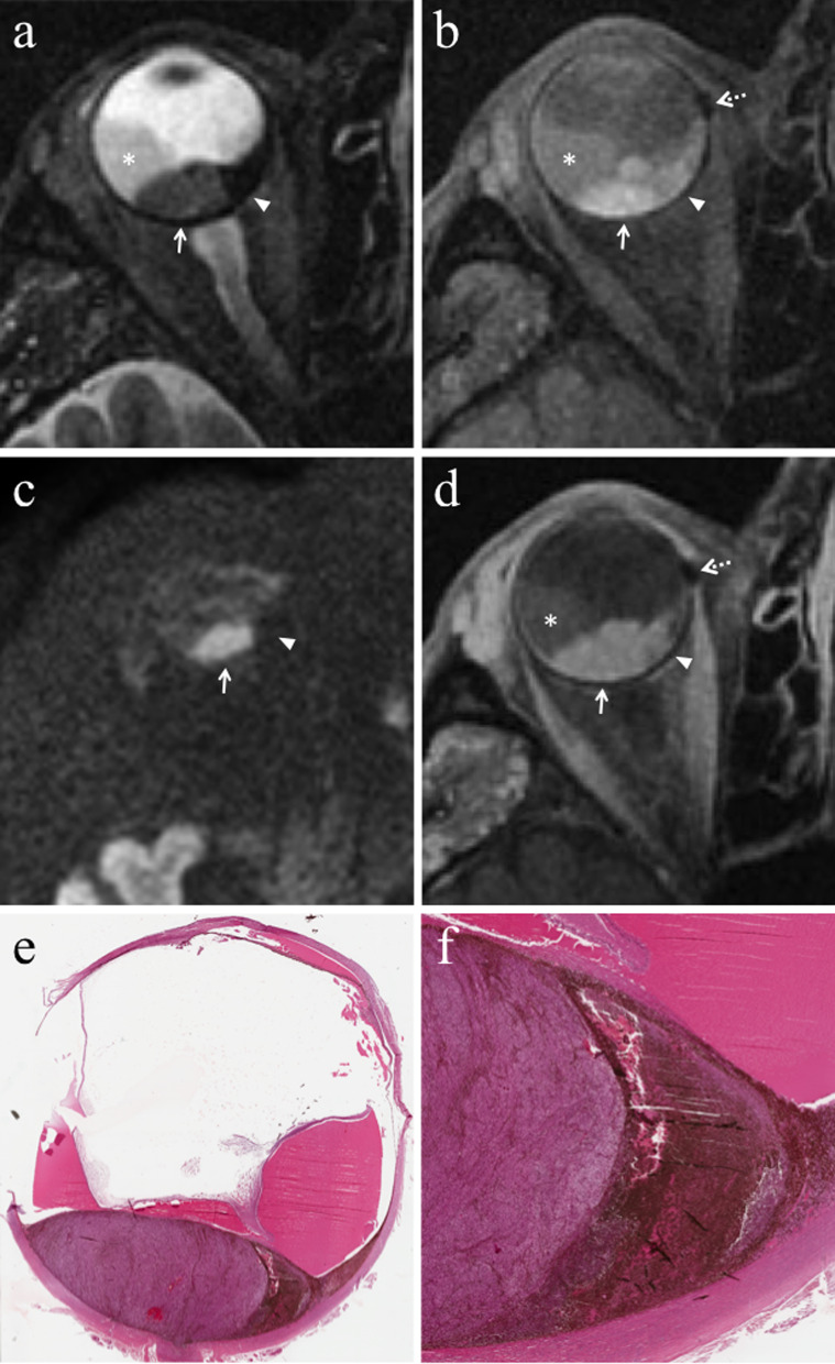
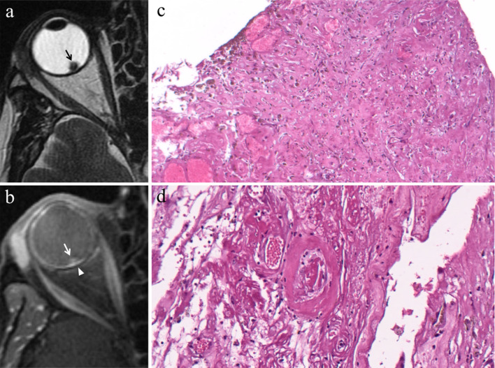
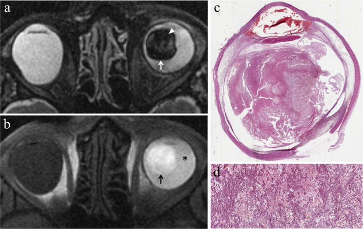
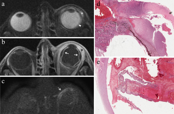
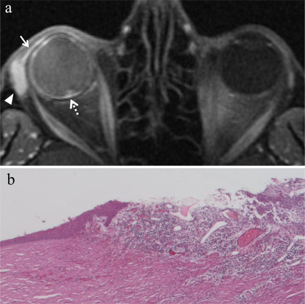
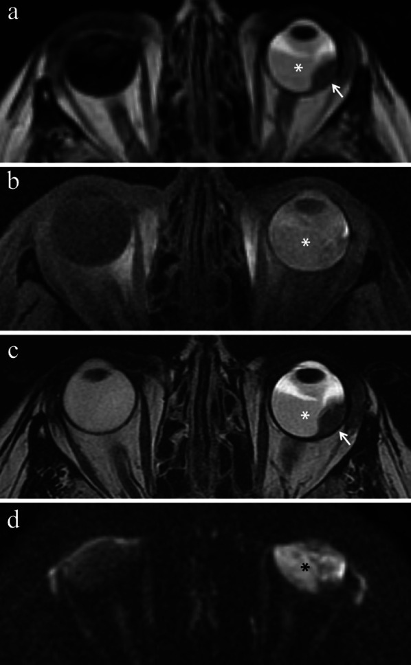
Therapy of uveal melanoma aims to preserve the eye and its function and to avoid metastatic dissemination. The treatment choice is difficult and must keep into account several factors; the therapeutic strategy of uveal melanoma should therefore be personalized, sometimes requiring to combine different treatment techniques. Nowadays globe-sparing radiotherapy techniques are often preferred to enucleation. Plaque brachytherapy, the most commonly used eye-preserving therapy, is suitable for small- and medium-sized uveal melanomas. Proton beam radiotherapy is indicated for tumours with noticeable size, challenging shape and location, but is more expensive and less available than brachytherapy. Enucleation is currently restricted to advanced tumours, uveal melanomas with orbital or optic nerve involvement, blind and painful eyes because of treatment-related complications (neovascular glaucoma, chronic inflammatory processes). The effect of proton beam therapy on neoplastic tissue is related to direct cytotoxic action of the radiations, impairment of neoplastic vascular supply and immunologic response. Complications after radiotherapy are frequent and numerous and mainly related to tumour thickness, radiation dose and distance between the tumour and optic nerve. The purpose of this pictorial review is to provide the radiologists with awareness about diagnostic methods and therapeutic options of uveal melanoma. In the present second section, we discuss the therapeutic management of uveal melanoma, describing the main ocular-conserving radiotherapic techniques. We subsequently present an overview of the effects of radiations on neoplastic tissue. Lastly, we review ocular complications following radiotherapy that should be evaluated by radiologists during follow-up MRI examinations.
葡萄膜黑色素瘤的治疗旨在保留眼球及其功能,并避免转移扩散。治疗选择颇具难度,必须考虑多个因素;因此,葡萄膜黑色素瘤的治疗策略应个性化,有时需要联合不同的治疗技术。如今,保留眼球的放射治疗技术通常比眼球摘除术更受青睐。敷贴近距离放射治疗是最常用的保留眼球治疗方法,适用于中小型葡萄膜黑色素瘤。质子束放射治疗适用于体积较大、形状复杂且位置特殊的肿瘤,但比近距离放射治疗费用更高且应用较少。目前,眼球摘除术仅限于晚期肿瘤、累及眼眶或视神经的葡萄膜黑色素瘤,以及因治疗相关并发症(新生血管性青光眼、慢性炎症过程)导致失明和疼痛的眼球。质子束治疗对肿瘤组织的作用与辐射的直接细胞毒性作用、肿瘤血管供应受损及免疫反应有关。放射治疗后的并发症常见且多样,主要与肿瘤厚度、辐射剂量以及肿瘤与视神经之间的距离有关。本图文综述的目的是让放射科医生了解葡萄膜黑色素瘤的诊断方法和治疗选择。在本文的第二部分,我们讨论葡萄膜黑色素瘤的治疗管理,描述主要的保留眼球放射治疗技术。随后,我们概述辐射对肿瘤组织的影响。最后,我们回顾放射治疗后的眼部并发症,放射科医生在随访磁共振成像检查时应评估这些并发症。