Department of Radiology, the First Affiliated Hospital of Guangzhou University of Chinese Medicine.
The First Clinical Medical College of Guangzhou University of Chinese Medicine, Guangzhou, China.
Medicine (Baltimore). 2021 Jun 4;100(22):e26212. doi: 10.1097/MD.0000000000026212.
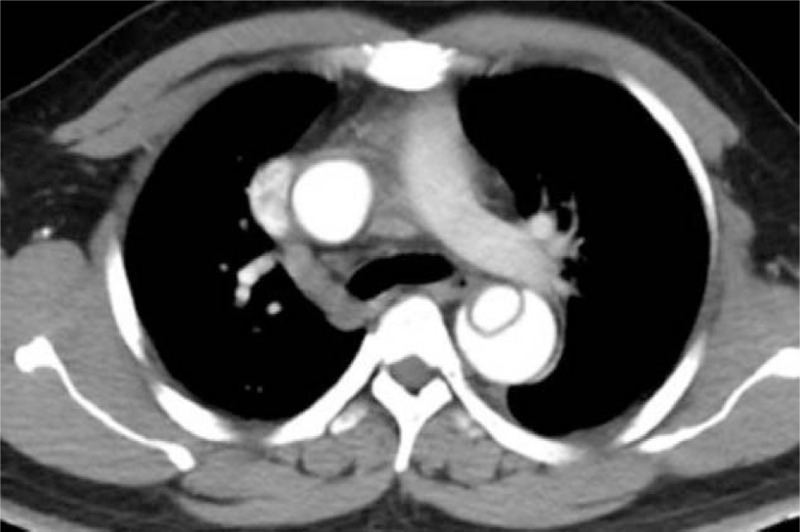
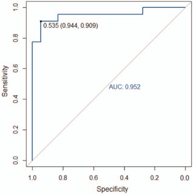
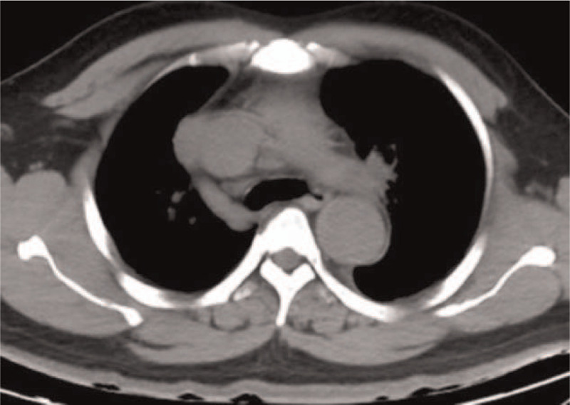
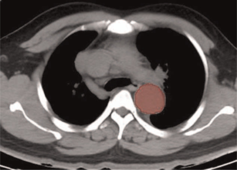
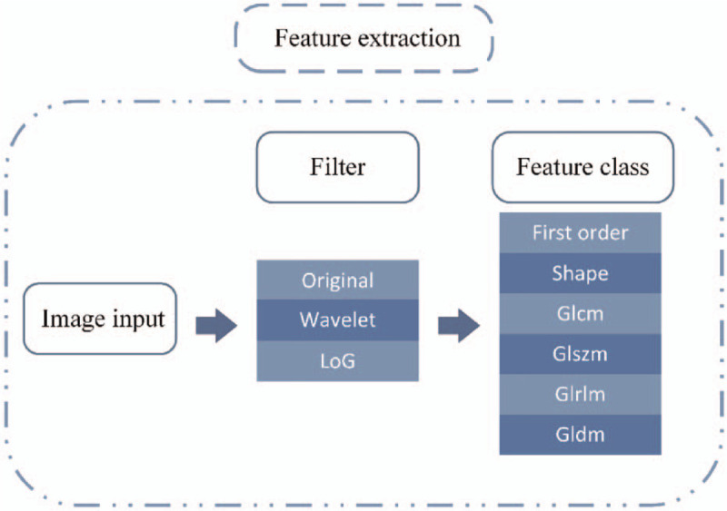
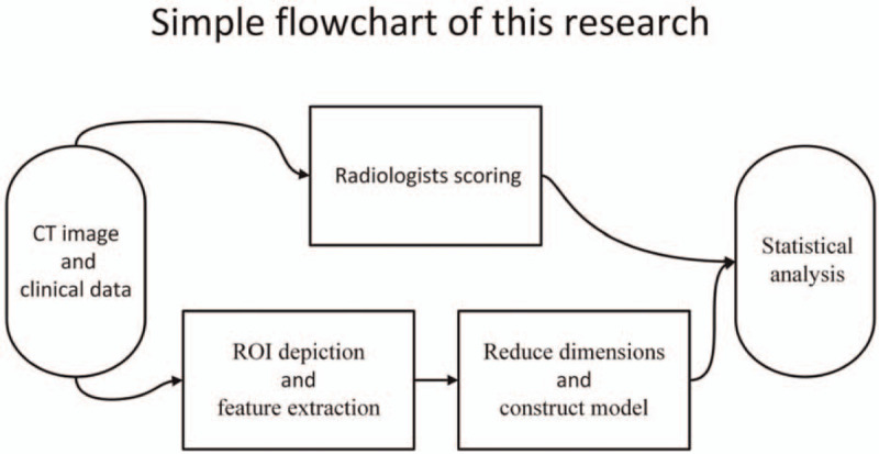
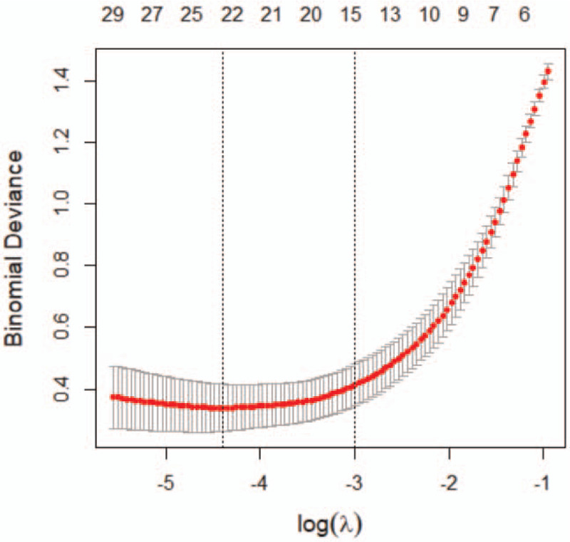
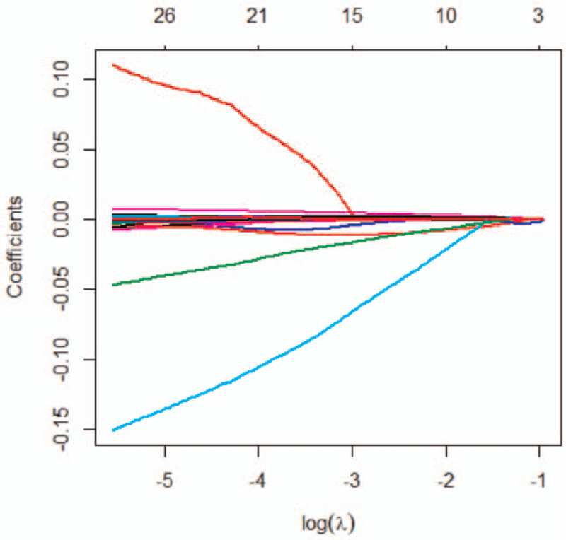
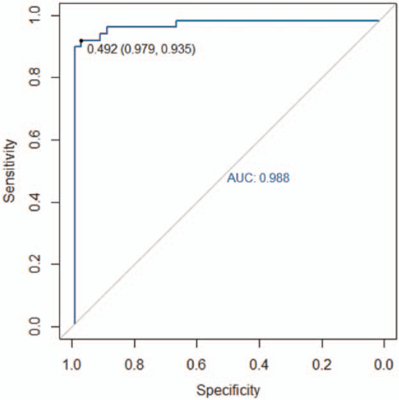
To investigate the diagnostic value of a computed tomography (CT) scan-based radiomics model for acute aortic dissection.For the dissection group, we retrospectively selected 50 patients clinically diagnosed with acute aortic dissection between October 2018 and November 2019, for whom non-contrast CT and CT angiography images were available. Fifty individuals with available non-contrast CT and CT angiography images for other causes were selected for inclusion in the non-dissection group. Based on the aortic dissection locations on the CT angiography images, we marked the corresponding regions-of-interest on the non-contrast CT images of both groups. We collected 1203 characteristic parameters from these regions by extracting radiomics features. Subsequently, we used a random number table to include 70 individuals in the training group and 30 in the validation group. Finally, we used the Lasso regression for dimension reduction and predictive model construction. The diagnostic performance of the model was evaluated by a receiver operating characteristic (ROC) curve.Fourteen characteristic parameters with non-zero coefficients were selected after dimension reduction. The accuracy, sensitivity, specificity, and area under the ROC curve of the prediction model for the training group were 94.3% (66/70), 91.2% (31/34), 97.2% (35/36), and 0.988 (95% confidence interval [CI]: 0.970-0.998), respectively. The respective values for the validation group were 90.0% (27/30), 94.1% (16/17), 84.6% (11/13), and 0.952 (95% CI: 0.883-0.986).Our non-contrast CT scan-based radiomics model accurately facilitated acute aortic dissection diagnosis.
探讨基于计算机断层扫描(CT)的放射组学模型对急性主动脉夹层的诊断价值。对于夹层组,我们回顾性选择了 2018 年 10 月至 2019 年 11 月期间临床上诊断为急性主动脉夹层的 50 例患者,这些患者有非对比 CT 和 CT 血管造影图像。选择了 50 名其他原因有非对比 CT 和 CT 血管造影图像的个体纳入非夹层组。基于 CT 血管造影图像上的主动脉夹层位置,我们在两组的非对比 CT 图像上标记了相应的感兴趣区域。我们从这些区域提取放射组学特征,收集了 1203 个特征参数。随后,我们使用随机数表将 70 人纳入训练组,30 人纳入验证组。最后,我们使用 Lasso 回归进行降维和预测模型构建。通过受试者工作特征(ROC)曲线评估模型的诊断性能。降维后选择了 14 个具有非零系数的特征参数。训练组预测模型的准确率、敏感度、特异度和 ROC 曲线下面积分别为 94.3%(66/70)、91.2%(31/34)、97.2%(35/36)和 0.988(95%置信区间[CI]:0.970-0.998)。验证组的相应值分别为 90.0%(27/30)、94.1%(16/17)、84.6%(11/13)和 0.952(95%CI:0.883-0.986)。我们的非对比 CT 扫描放射组学模型准确地促进了急性主动脉夹层的诊断。