Zhang Yuyanan, Liao Jinmin, Li Qianqian, Zhang Xiao, Liu Lijun, Yan Jun, Zhang Dai, Yan Hao, Yue Weihua
Institute of Mental Health, Peking University Sixth Hospital, Beijing, China.
Key Laboratory of Mental Health, Ministry of Health & National Clinical Research Center for Mental Disorders, Peking University, Beijing, China.
Front Psychiatry. 2021 May 21;12:681701. doi: 10.3389/fpsyt.2021.681701. eCollection 2021.
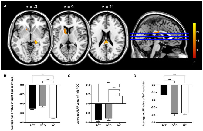
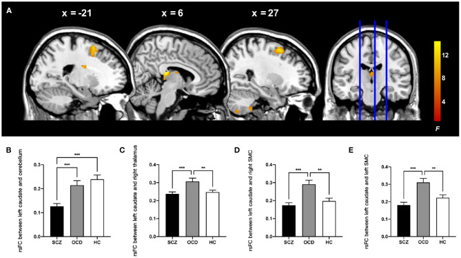
Schizophrenia (SCZ) and obsessive-compulsive disorder (OCD) are classified as two chronic psychiatric disorders with high comorbidity rate and shared clinical symptoms. Abnormal spontaneous brain activity within the cortical-striatal neural circuits has been observed in both disorders. However, it is unclear if the common or distinct neural abnormalities underlie the neurobiological substrates in the resting state. Resting-state fMRI data were collected from 88 patients with SCZ, 58 patients with OCD, and 72 healthy control subjects. First, we examined differences in amplitude of low-frequency fluctuations (ALFF) among three groups. Resting-state functional connectivity (rsFC) analysis with the brain region that showed different ALFF as the seed was then conducted to identify the changes in brain networks. Finally, we examined the correlation between the altered activities and clinical symptoms. Both the patients with SCZ and OCD showed increased ALFF in the right hippocampus and decreased ALFF in the left posterior cingulate cortex (PCC). SCZ patients exhibited increased ALFF in the left caudate [voxel-level family-wise error (FWE) < 0.05] and decreased rsFC between the left caudate and right cerebellum, which correlated with positive symptoms. The left caudate showed increased rsFC with the right thalamus and bilateral supplementary motor complex (SMC) in OCD patients (cluster-level FWE < 0.05). The hippocampus and PCC are common regions presenting abnormal local spontaneous neuronal activities in both SCZ and OCD, while the abnormality of the striatum can reflect the differences. Increased ALFF in the striatum and symptom-related weakened rsFC between the caudate and cerebellum showed SCZ specificity. Enhanced rsFC between the caudate and SMC may be a key characteristic in OCD. Our research shows the similarities and differences between the two diseases from the perspective of resting-state fMRI, which provides clues to understand the disease and find methods for treatment.
精神分裂症(SCZ)和强迫症(OCD)被归类为两种慢性精神疾病,共病率高且有共同的临床症状。在这两种疾病中均观察到皮质-纹状体神经回路内的异常自发脑活动。然而,尚不清楚静息状态下的神经生物学基础是由共同的还是独特的神经异常所导致。我们收集了88例SCZ患者、58例OCD患者和72名健康对照者的静息态功能磁共振成像(fMRI)数据。首先,我们检查了三组之间低频波动幅度(ALFF)的差异。然后以显示出不同ALFF的脑区为种子进行静息态功能连接(rsFC)分析,以识别脑网络的变化。最后,我们检查了活动改变与临床症状之间的相关性。SCZ患者和OCD患者在右侧海马体中均表现出ALFF增加,而在左侧后扣带回皮质(PCC)中ALFF降低。SCZ患者左侧尾状核的ALFF增加[体素水平家族性错误率(FWE)<0.05],并且左侧尾状核与右侧小脑之间的rsFC降低,这与阳性症状相关。在OCD患者中,左侧尾状核与右侧丘脑和双侧辅助运动复合体(SMC)之间的rsFC增加(簇水平FWE<0.05)。海马体和PCC是SCZ和OCD中均出现局部自发神经元活动异常的共同区域,而纹状体的异常可以反映差异。纹状体中ALFF增加以及尾状核与小脑之间与症状相关的rsFC减弱显示出SCZ的特异性。尾状核与SMC之间增强的rsFC可能是OCD的一个关键特征。我们的研究从静息态fMRI的角度显示了这两种疾病之间的异同,为理解疾病和寻找治疗方法提供了线索。