From the Department of Neurology (T.F., Y.M., R.Y., K.I., A.S., J.-i.K.), Neurological Institute, Graduate School of Medical Sciences, Kyushu University, Fukuoka, Japan; Department of Neurology (E.-J.L., Y.-M.L., K.-K.K.), Asan Medical Center, University of Ulsan, College of Medicine, Seoul, South Korea; Translational Neuroscience Center (J.-i.K.), Graduate School of Medicine, and School of Pharmacy at Fukuoka, International University of Health and Welfare, Okawa; and Department of Neurology (J.-i.K.), Brain and Nerve Center, Fukuoka Central Hospital, International University of Health and Welfare, Japan.
Neurol Neuroimmunol Neuroinflamm. 2021 Jun 7;8(5). doi: 10.1212/NXI.0000000000001028. Print 2021 Jul.
To assess the prevalence of antiplexin D1 antibodies (plexin D1-immunoglobulin G [IgG]) in small fiber neuropathy (SFN) and the effects of these antibodies in vivo.
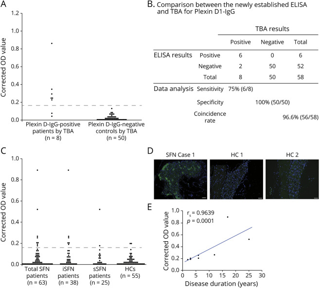
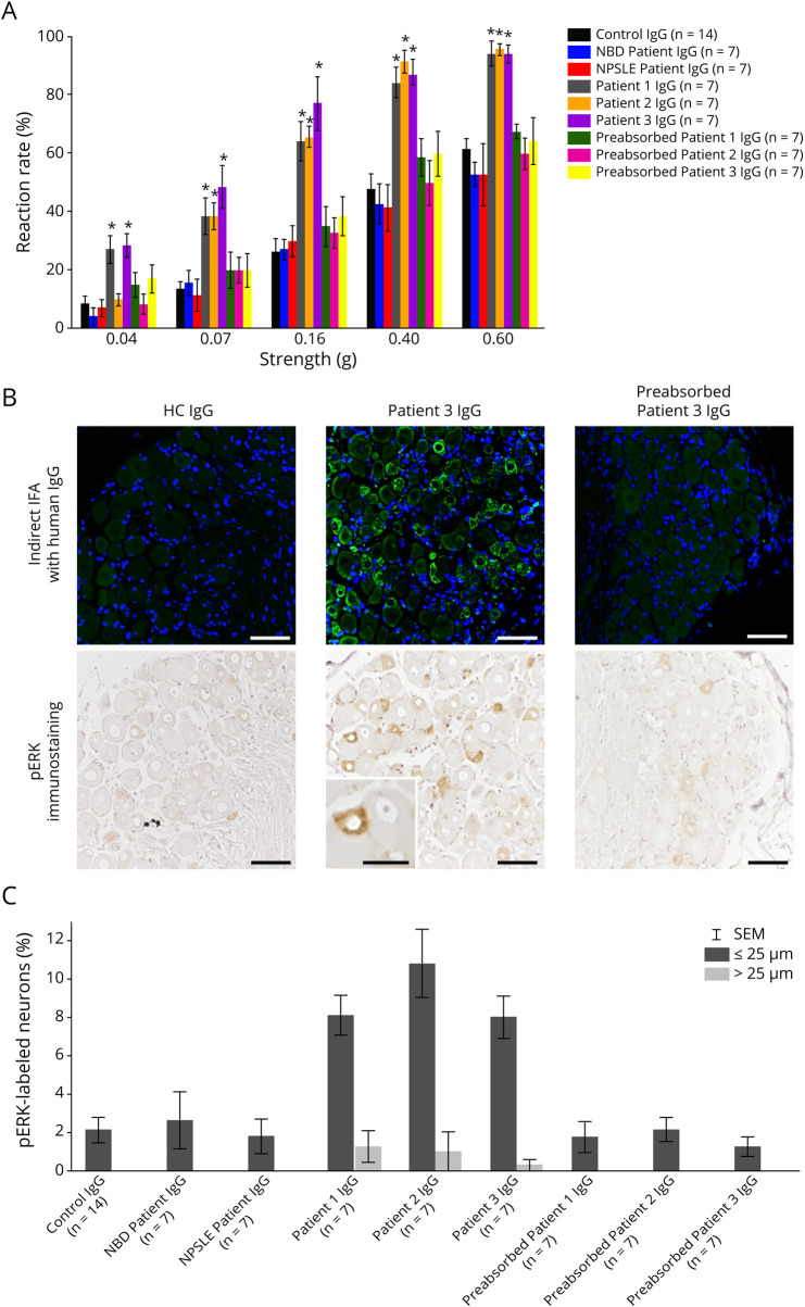
We developed an ELISA for plexin D1-IgG using a recombinant extracellular domain of human plexin D1 containing the major epitope and sera from 58 subjects previously studied with a standard tissue-based indirect immunofluorescence assay (TBA). We screened 63 patients with probable SFN and 55 healthy controls (HCs) for serum plexin D1-IgG using ELISA. The results were confirmed by TBA. IgG from 3 plexin D1-IgG-positive patients, 2 plexin D1-IgG-negative inflammatory disease controls, and 2 HCs was intrathecally injected into mice, which were assessed for mechanical and thermal hypersensitivity 24 and 48 hours after injection.
The ELISA had 75% sensitivity and 100% specificity using the TBA as a standard, and the coincidence rate of ELISA to TBA was 96.6% (56/58). The frequency of plexin D1-IgG was higher in patients with SFN than in HCs (12.7% [8/63] vs 0.0% [0/55], = 0.007). Purified IgG from all 3 plexin D1-IgG-positive patients, but not 2 plexin D1-IgG-negative patients, induced significant mechanical and/or thermal hypersensitivity compared with IgG from HCs. In mice injected with plexin D1-IgG-positive but not D1-IgG-negative patient IgG, phosphorylated extracellular signal-regulated protein kinase immunoreactivity, an activation marker, was confined to small dorsal root ganglion neurons and was significantly more abundant than in mice injected with HC IgG.
Plexin D1-IgG is pathogenic but with low prevalence and is a potential biomarker for immunotherapy in SFN.
评估抗多聚蛋白 D1 抗体(plexin D1-免疫球蛋白 G [IgG])在小纤维神经病(SFN)中的流行率以及这些抗体在体内的作用。
我们使用包含主要表位的人多聚蛋白 D1 的重组细胞外结构域开发了用于 plexin D1-IgG 的 ELISA,并使用先前使用标准基于组织的间接免疫荧光测定法(TBA)进行研究的 58 名受试者的血清。我们使用 ELISA 筛查了 63 例疑似 SFN 患者和 55 名健康对照者(HCs)的血清 plexin D1-IgG。通过 TBA 确认结果。将来自 3 例 plexin D1-IgG 阳性患者、2 例 plexin D1-IgG 阴性炎性疾病对照者和 2 例 HCs 的 IgG 鞘内注射到小鼠中,在注射后 24 和 48 小时评估它们的机械和热敏感性。
使用 TBA 作为标准,ELISA 的灵敏度为 75%,特异性为 100%,ELISA 与 TBA 的符合率为 96.6%(56/58)。SFN 患者 plexin D1-IgG 的频率高于 HCs(12.7%[8/63] vs 0.0%[0/55], = 0.007)。来自所有 3 例 plexin D1-IgG 阳性患者的纯化 IgG,但不是来自 2 例 plexin D1-IgG 阴性患者的 IgG,与来自 HCs 的 IgG 相比,可诱导明显的机械和/或热敏感性。在注射了 plexin D1-IgG 阳性但不是 D1-IgG 阴性患者 IgG 的小鼠中,磷酸化细胞外信号调节蛋白激酶免疫反应性,一种激活标志物,局限于小的背根神经节神经元,并且比注射了 HC IgG 的小鼠中的更丰富。
plexin D1-IgG 是致病性的,但患病率较低,是 SFN 免疫治疗的潜在生物标志物。