Department of Biological and Biochemical Sciences, Faculty of Chemical Technology, University of Pardubice, Studentska 573, 532 10, Pardubice, Czech Republic.
Sci Rep. 2021 Jun 7;11(1):11921. doi: 10.1038/s41598-021-91380-3.
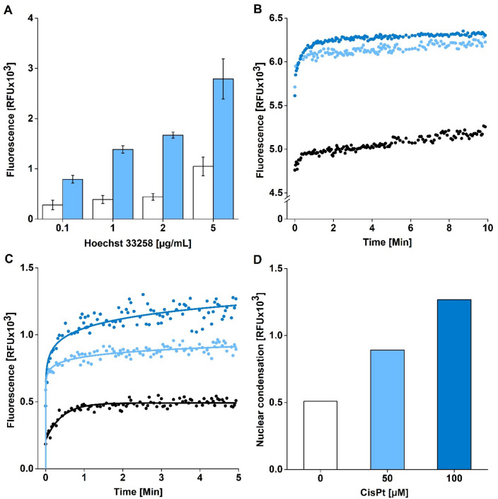
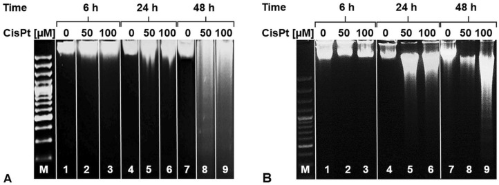
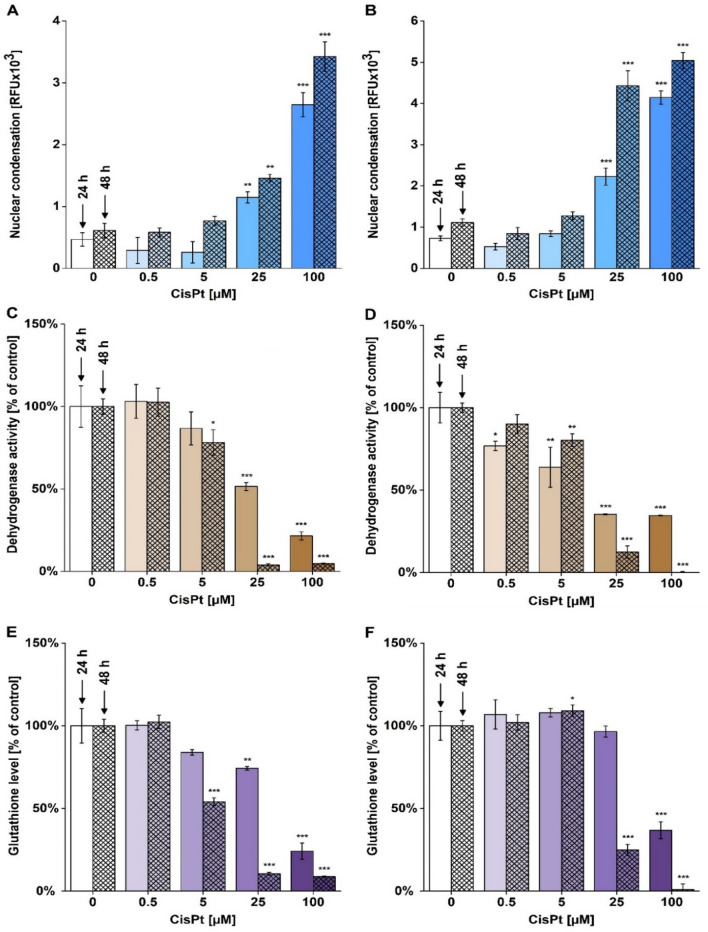
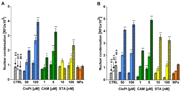
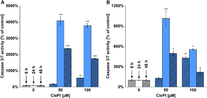
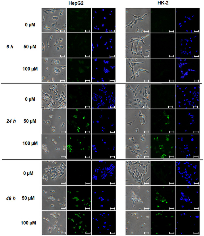
At present, nuclear condensation and fragmentation have been estimated also using Hoechst probes in fluorescence microscopy and flow cytometry. However, none of the methods used the Hoechst probes for quantitative spectrofluorometric assessment. Therefore, the aim of the present study was to develop a spectrofluorometric assay for detection of nuclear condensation and fragmentation in the intact cells. We used human hepatoma HepG2 and renal HK-2 cells cultured in 96-well plates treated with potent apoptotic inducers (i.e. cisplatin, staurosporine, camptothecin) for 6-48 h. Afterwards, the cells were incubated with Hoechst 33258 (2 µg/mL) and the increase of fluorescence after binding of the dye to DNA was measured. The developed spectrofluorometric assay was capable to detect nuclear changes caused by all tested apoptotic inducers. Then, we compared the outcomes of the spectrofluorometric assay with other methods detecting cell impairment and apoptosis (i.e. WST-1 and glutathione tests, TUNEL, DNA ladder, caspase activity, PARP-1 and JNKs expressions). We found that our developed spectrofluorometric assay provided results of the same sensitivity as the TUNEL assay but with the advantages of being fast processing, low-cost and a high throughput. Because nuclear condensation and fragmentation can be typical markers of cell death, especially in apoptosis, we suppose that the spectrofluorometric assay could become a routinely used method for characterizing cell death processes.
目前,也可以使用荧光显微镜和流式细胞术中的 Hoechst 探针来估计核浓缩和核碎裂。然而,目前使用 Hoechst 探针进行定量分光荧光评估的方法尚未建立。因此,本研究旨在开发一种用于检测完整细胞中核浓缩和核碎裂的分光荧光分析方法。我们使用人肝癌 HepG2 和肾 HK-2 细胞,在 96 孔板中培养,用有效的凋亡诱导剂(即顺铂、星形孢菌素、喜树碱)处理 6-48 小时。然后,用 Hoechst 33258(2μg/mL)孵育细胞,并测量染料与 DNA 结合后荧光的增加。开发的分光荧光分析方法能够检测所有测试的凋亡诱导剂引起的核变化。然后,我们将分光荧光分析的结果与其他检测细胞损伤和凋亡的方法(即 WST-1 和谷胱甘肽测定法、TUNEL、DNA 梯、半胱天冬酶活性、PARP-1 和 JNKs 表达)进行了比较。我们发现,我们开发的分光荧光分析方法与 TUNEL 测定法具有相同的敏感性,但具有快速处理、低成本和高通量的优势。因为核浓缩和核碎裂可能是细胞死亡的典型标志物,特别是在凋亡中,我们推测分光荧光分析方法可能成为一种常规的细胞死亡过程的特征方法。