Department of Translational Medical Sciences, Section of Pediatrics, University of Naples Federico II, Naples, Italy.
Department of Advanced Biomedical Sciences, Pathology Unit, University of Naples Federico II, Naples, Italy.
JAMA Netw Open. 2021 Jun 1;4(6):e2111369. doi: 10.1001/jamanetworkopen.2021.11369.
Chilblain-like lesions have been one of the most frequently described cutaneous manifestations during the COVID-19 pandemic. Their etiopathogenesis, including the role of SARS-CoV-2, remains elusive.
To examine the association of chilblain-like lesions with SARS-CoV-2 infection.
DESIGN, SETTING, AND PARTICIPANTS: This prospective case series enrolled 17 adolescents who presented with chilblain-like lesions from April 1 to June 30, 2020, at a tertiary referral academic hospital in Italy.
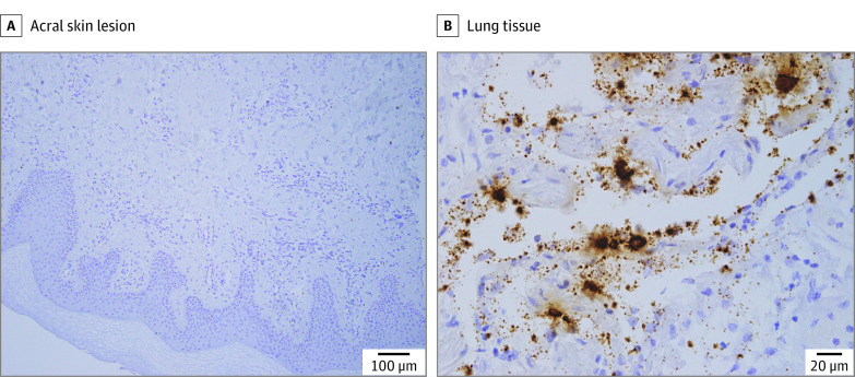
Macroscopic (clinical and dermoscopic) and microscopic (histopathologic) analysis contributed to a thorough understanding of the lesions. Nasopharyngeal swab, serologic testing, and in situ hybridization of the skin biopsy specimens were performed to test for SARS-CoV-2 infection. Laboratory tests explored signs of systemic inflammation or thrombophilia. Structural changes in peripheral microcirculation were investigated by capillaroscopy.
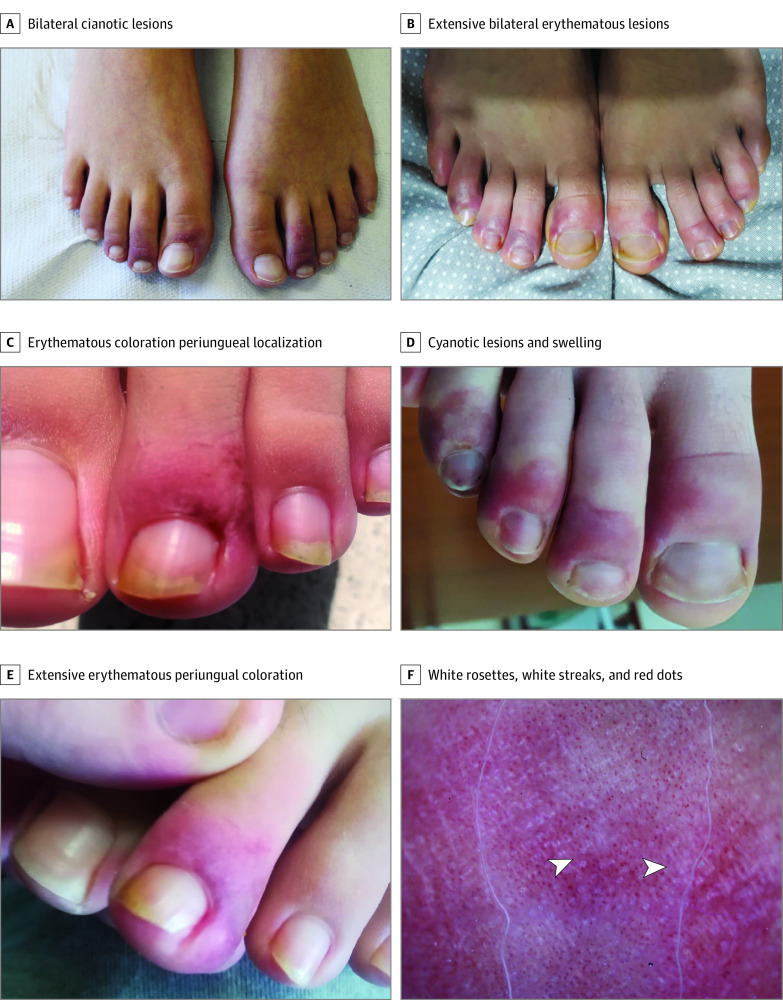
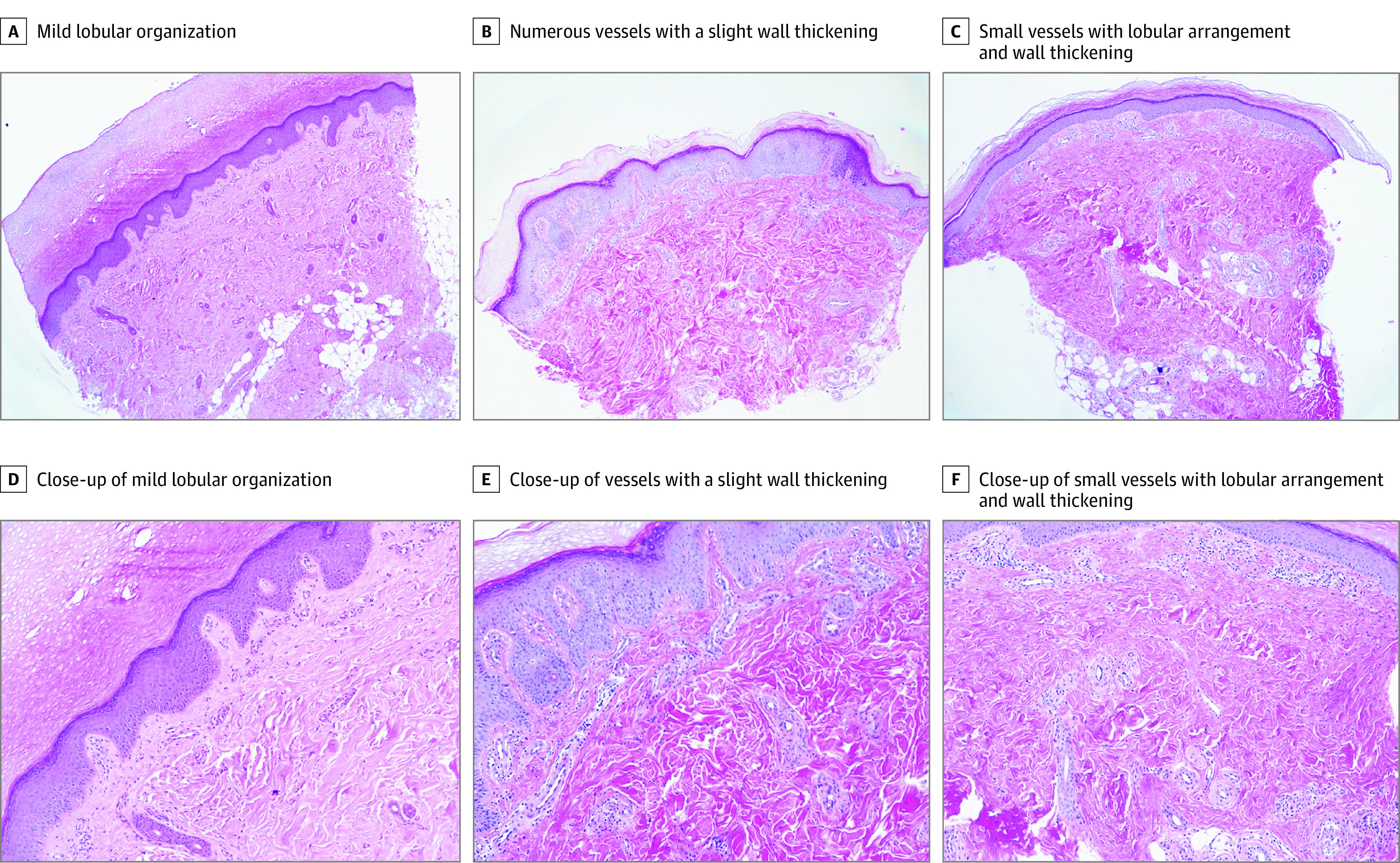
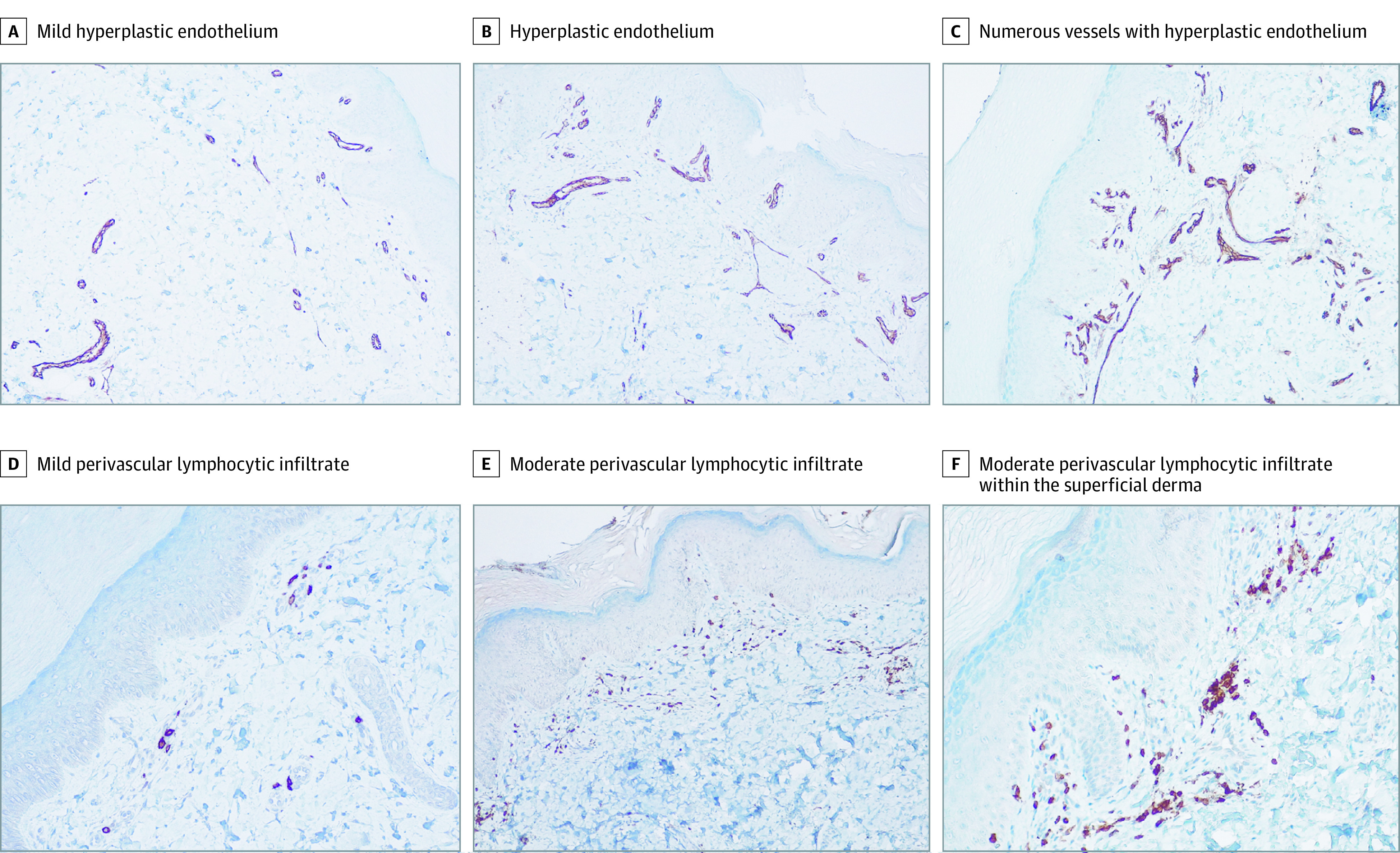
Of the 17 adolescents (9 [52.9%] male; median [interquartile range] age, 13.2 [12.5-14.3] years) enrolled during the first wave of the COVID-19 pandemic, 16 (94.1%) had bilaterally localized distal erythematous or cyanotic lesions. A triad of red dots (16 [100%]), white rosettes (11 [68.8%]), and white streaks (10 [62.5%]) characterized the dermoscopic picture. Histologic analysis revealed a remodeling of the dermal blood vessels with a lobular arrangement, wall thickening, and a mild perivascular lymphocytic infiltrate. SARS-CoV-2 infection was excluded by molecular and serologic testing. In situ hybridization did not highlight the viral genome in the lesions.
This study delineated the clinical, histologic, and laboratory features of chilblain-like lesions that emerged during the COVID-19 pandemic, and its findings do not support their association with SARS-CoV-2 infection. The lesions occurred in otherwise healthy adolescents, had a long but benign course to self-resolution, and were characterized by a microvascular remodeling with perivascular lymphocytic infiltrate but no other signs of vasculitis. These results suggest that chilblain-like lesions do not imply a concomitant SARS-CoV-2 infection. Ongoing studies will help clarify the etiopathogenic mechanisms.
冻疮样病变是 COVID-19 大流行期间最常描述的皮肤表现之一。其发病机制,包括 SARS-CoV-2 的作用,仍然难以捉摸。
研究冻疮样病变与 SARS-CoV-2 感染的相关性。
设计、地点和参与者:这项前瞻性病例系列研究纳入了 2020 年 4 月 1 日至 6 月 30 日期间在意大利一家三级转诊学术医院就诊的 17 名出现冻疮样病变的青少年。
进行了宏观(临床和皮肤镜)和微观(组织病理学)分析,以全面了解病变。进行了鼻咽拭子、血清学检测和皮肤活检标本的原位杂交,以检测 SARS-CoV-2 感染。实验室检测探索了全身炎症或血栓形成的迹象。通过毛细血管镜检查研究了外周微循环的结构变化。
在 COVID-19 大流行的第一波期间,共纳入 17 名青少年(9 名[52.9%]为男性;中位[四分位间距]年龄为 13.2[12.5-14.3]岁),16 名(94.1%)存在双侧局灶性远端红斑或青紫色病变。红色小点三联征(16 名[100%])、白色罗勒(11 名[68.8%])和白色条纹(10 名[62.5%])是皮肤镜下的特征性表现。组织学分析显示真皮血管重塑,呈小叶状排列,壁增厚,轻度血管周围淋巴细胞浸润。通过分子和血清学检测排除了 SARS-CoV-2 感染。原位杂交未在病变中突出显示病毒基因组。
本研究描绘了 COVID-19 大流行期间出现的冻疮样病变的临床、组织学和实验室特征,其研究结果不支持它们与 SARS-CoV-2 感染相关。病变发生在其他健康的青少年中,具有较长但良性的自限性病程,其特征是血管周围淋巴细胞浸润但没有其他血管炎表现的微血管重塑。这些结果表明,冻疮样病变并不意味着同时存在 SARS-CoV-2 感染。正在进行的研究将有助于阐明发病机制。