He Shanqing, Wang Youcheng, Yao Yajun, Cao Zhen, Yin Junkui, Zi Liuliu, Chen Huiyu, Fu Yuntao, Wang Xi, Zhao Qingyan
Department of Cardiology, Renmin Hospital of Wuhan University, Wuhan, China.
Cardiovascular Research Institute of Wuhan University, Wuhan, China.
Front Cardiovasc Med. 2021 May 31;8:656631. doi: 10.3389/fcvm.2021.656631. eCollection 2021.
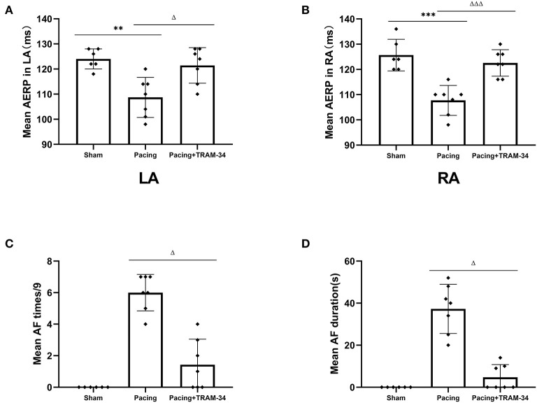
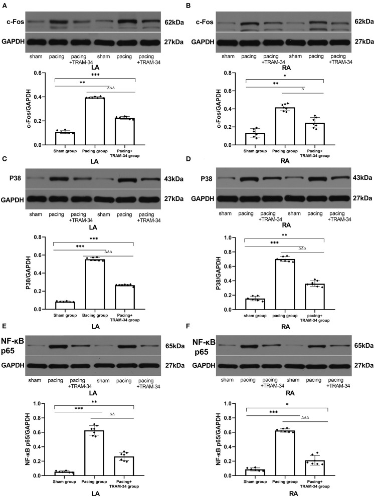
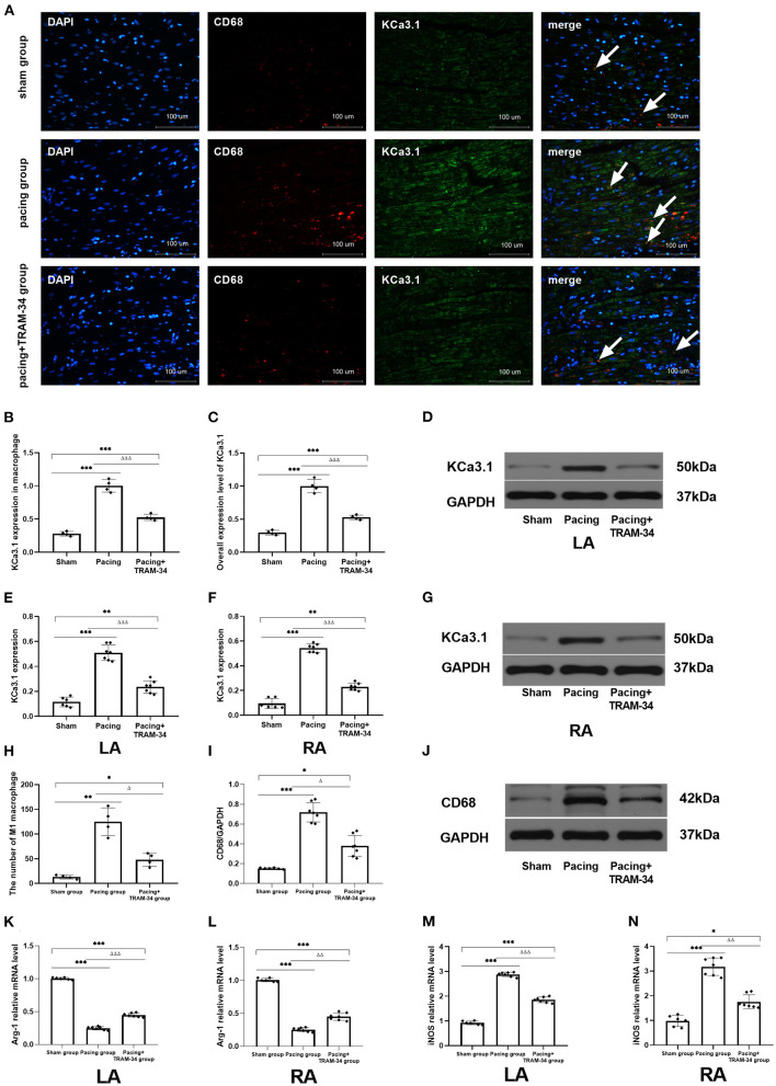
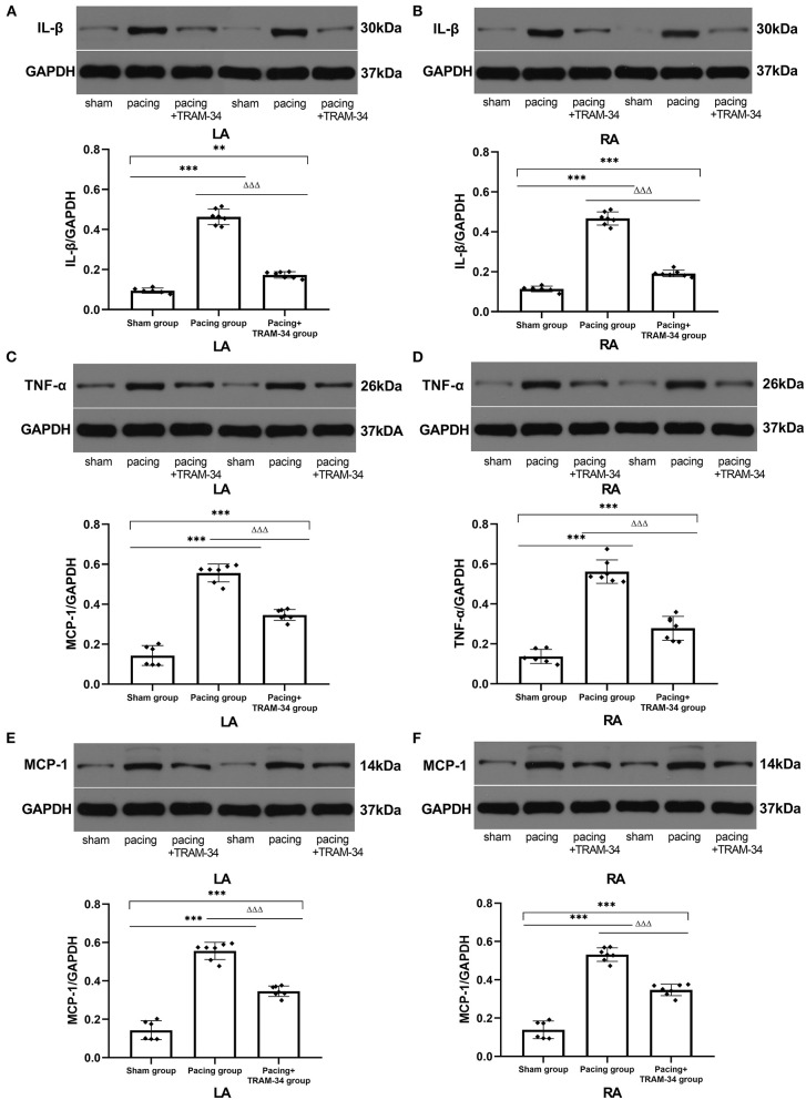
To investigate the role of KCa3. 1 inhibition in macrophage pro-inflammatory polarization and vulnerability to atrial fibrillation (AF) in a canine model with prolonged rapid atrial pacing. Twenty beagle dogs (weighing 8-10 kg) were randomly assigned to a sham group ( = 6), pacing group ( = 7) and pacing+TRAM-34 group ( = 7). An experimental model of AF was established by rapid pacing. TRAM-34 was administered to the Pacing+TRAM-34 group by slow intravenous injection (10 mg/kg), 3 times each day. After 7 days of pacing, the electrophysiology was measured . The levels of interleukin-1β (IL-1β), monocyte chemotactic protein-1 (MCP-1), tumor necrosis factor-α (TNF-α), CD68, c-Fos, p38, and NF-κB p65 in both atriums were measured by Western blotting, and the levels of inducible nitric oxide synthase (iNOS) and arginase1 (Arg-1) were measured by real-time PCR. Macrophage and KCa3.1 in macrophage in the atrium were quantized following double labeled immunofluorescent. Greater inducibility of AF, an extended duration of AF and lower atrial effective refractory period (AERP) were observed in the pacing group compared with those in the sham group. Both CD68-labeled macrophage and the expression of KCa3.1 in macrophage were elevated in the pacing group and inhibited by TRAM-34, led to higher iNOS expression, lower Arg-1 expression, elevated levels of IL-1β, MCP-1, and TNF-α in the atria, which could be reversed by TRAM-34 treatment (all < 0.01). KCa3.1 channels were possibly activated via the p38/AP-1/NF-κB signaling pathway. Inhibition of KCa3.1 suppresses vulnerability to AF by attenuating macrophage pro-inflammatory polarization and inflammatory cytokine secretion in a canine model with prolonged rapid atrial pacing.
为研究在犬类长时间快速心房起搏模型中,抑制大电导钙激活钾通道3.1(KCa3.1)对巨噬细胞促炎极化及心房颤动(AF)易感性的作用。将20只比格犬(体重8 - 10千克)随机分为假手术组(n = 6)、起搏组(n = 7)和起搏+TRAM - 34组(n = 7)。通过快速起搏建立AF实验模型。起搏+TRAM - 34组通过缓慢静脉注射(10毫克/千克)给予TRAM - 34,每日3次。起搏7天后,测量电生理指标。通过蛋白质免疫印迹法测量两心房中白细胞介素 - 1β(IL - 1β)、单核细胞趋化蛋白 - 1(MCP - 1)、肿瘤坏死因子 - α(TNF - α)、CD68、c - Fos、p38和核因子κB p65(NF - κB p65)的水平,通过实时聚合酶链反应测量诱导型一氧化氮合酶(iNOS)和精氨酸酶1(Arg - 1)的水平。采用双标免疫荧光法定量心房中的巨噬细胞及巨噬细胞中的KCa3.1。与假手术组相比,起搏组AF的诱导性更高、AF持续时间更长且心房有效不应期(AERP)更低。起搏组中CD68标记的巨噬细胞及巨噬细胞中KCa3.1的表达均升高,且被TRAM - 34抑制,导致心房中iNOS表达升高、Arg - 1表达降低、IL - 1β、MCP - 1和TNF - α水平升高,而TRAM - 34治疗可使其逆转(均P < 0.01)。KCa3.1通道可能通过p38/激活蛋白 - 1(AP - 1)/NF - κB信号通路被激活。在犬类长时间快速心房起搏模型中,抑制KCa3.1可通过减弱巨噬细胞促炎极化及炎性细胞因子分泌来抑制AF易感性。