Department of Ophthalmology, Korea University College of Medicine, Seoul, Korea.
Korean J Ophthalmol. 2021 Aug;35(4):311-317. doi: 10.3341/kjo.2021.0007. Epub 2021 Jun 21.
To evaluate the effects of baseline trabecular meshwork (TM) and Schlemm's canal (SC) microstructures on intraocular pressure (IOP) reduction amount in treatment-naïve patients with primary open-angle glaucoma (POAG).
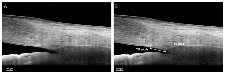
A total 69 eyes of POAG patients who had not been treated with IOP-lowering agent were enrolled in this retrospective study. The patients had been prescribed topical IOP-lowering agent and used it for 1 year. The morphologic features of the TM and SC were collected using anterior segment module of spectral-domain optical coherence tomography with enhanced depth imaging at baseline. Images of the nasal and temporal corneoscleral limbus were obtained with serial horizontal enhanced depth imaging B-scans and TM width and SC area were measured in each scan. We investigated the effects of baseline TM and SC microstructures on IOP reduction amount.
The baseline IOP of 69 glaucomatous eyes was 17.9 ± 3.8 mmHg, and the mean amount of IOP reduction was 3.5 ± 2.1 mmHg after 1 year. Mean TM widths of nasal and temporal sector were 470.33 ± 80.05 and 479.74 ± 79.59 μm, respectively. SC area was measured as 4,818.50 ± 1,464.28, 4,604.23 ± 1,567.73 μm2 at nasal sector and temporal sector, respectively. The correlation analysis revealed a positive correlation between SC area and average amount of IOP reduction, indicating that the larger baseline SC area, the greater the IOP drop with topical IOP-lowering agents. However, no correlation was found between TM width and IOP lowering amount in patients with POAG.
The baseline SC area showed positive correlation with the IOP reduction amount in patients with POAG. This finding suggests that the SC area can be a clinical parameter to predict the IOP reduction amount before using IOP-lowering agents in POAG patient.
评估原发性开角型青光眼(POAG)治疗初治患者的基线小梁网(TM)和施莱姆管(SC)微观结构对眼压(IOP)降低量的影响。
本回顾性研究纳入了 69 只未经降眼压药物治疗的 POAG 患者的眼睛。这些患者已被处方局部降眼压药物,并使用了 1 年。在基线时,使用带有增强深度成像的频域光学相干断层扫描的前段模块收集 TM 和 SC 的形态特征。使用连续的水平增强深度成像 B 扫描获得鼻侧和颞侧角巩膜缘的图像,并在每个扫描中测量 TM 宽度和 SC 面积。我们研究了基线 TM 和 SC 微观结构对 IOP 降低量的影响。
69 只青光眼眼中的基线 IOP 为 17.9 ± 3.8 mmHg,1 年后平均 IOP 降低量为 3.5 ± 2.1 mmHg。鼻侧和颞侧的平均 TM 宽度分别为 470.33 ± 80.05 μm 和 479.74 ± 79.59 μm。SC 面积在鼻侧和颞侧分别测量为 4,818.50 ± 1,464.28 μm2和 4,604.23 ± 1,567.73 μm2。相关分析显示 SC 面积与平均 IOP 降低量呈正相关,表明基线 SC 面积越大,局部降眼压药物的眼压降幅越大。然而,在 POAG 患者中,TM 宽度与 IOP 降低量之间未发现相关性。
POAG 患者的基线 SC 面积与 IOP 降低量呈正相关。这一发现表明,在 POAG 患者使用降眼压药物之前,SC 面积可以作为预测 IOP 降低量的临床参数。