• 文献检索
  • 文档翻译
  • 深度研究
  • 学术资讯
  • Suppr Zotero 插件Zotero 插件
  • 邀请有礼
  • 套餐&价格
  • 历史记录
应用&插件
Suppr Zotero 插件Zotero 插件浏览器插件Mac 客户端Windows 客户端微信小程序
定价
高级版会员购买积分包购买API积分包
服务
文献检索文档翻译深度研究API 文档MCP 服务
关于我们
关于 Suppr公司介绍联系我们用户协议隐私条款
关注我们

Suppr 超能文献

核心技术专利:CN118964589B侵权必究
粤ICP备2023148730 号-1Suppr @ 2026

文献检索

告别复杂PubMed语法,用中文像聊天一样搜索,搜遍4000万医学文献。AI智能推荐,让科研检索更轻松。

立即免费搜索

文件翻译

保留排版,准确专业,支持PDF/Word/PPT等文件格式,支持 12+语言互译。

免费翻译文档

深度研究

AI帮你快速写综述,25分钟生成高质量综述,智能提取关键信息,辅助科研写作。

立即免费体验

叉头框蛋白 C2 在青光眼病理中的小梁网和施莱姆氏管中的差异作用。

Differential roles of FOXC2 in the trabecular meshwork and Schlemm's canal in glaucomatous pathology.

机构信息

Feinberg Cardiovascular and Renal Research Institute, Department of Medicine, Feinberg School of Medicine, Northwestern University, Chicago, IL, USA.

Department of Biomedical Engineering, Northwestern University, Evanston, IL, USA.

出版信息

Life Sci Alliance. 2023 Jul 6;6(9). doi: 10.26508/lsa.202201721. Print 2023 Sep.

DOI:10.26508/lsa.202201721
PMID:37414529
原文链接:https://pmc.ncbi.nlm.nih.gov/articles/PMC10326420/
Abstract

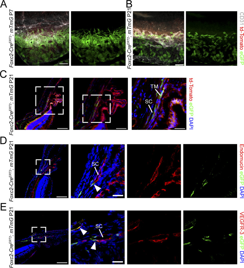
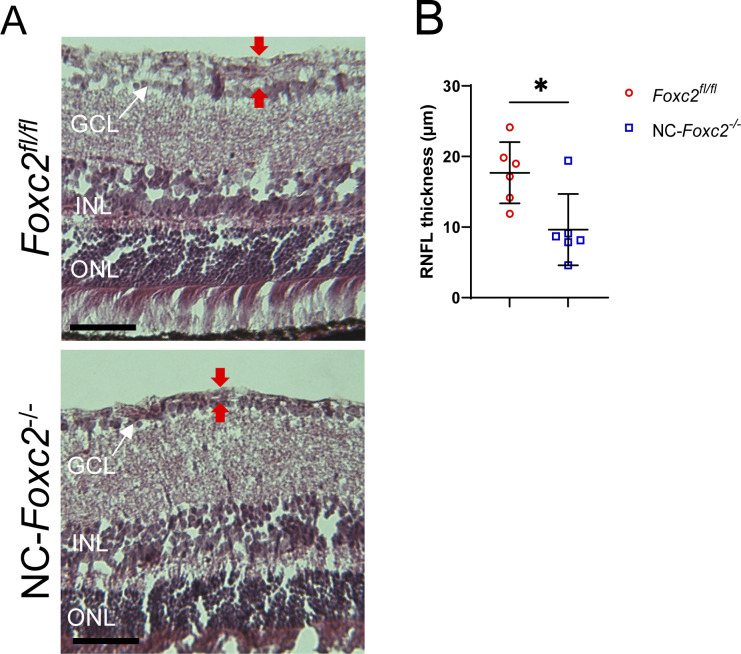
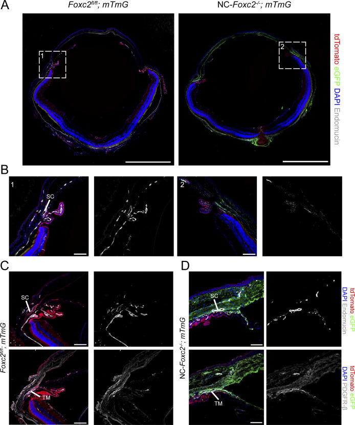
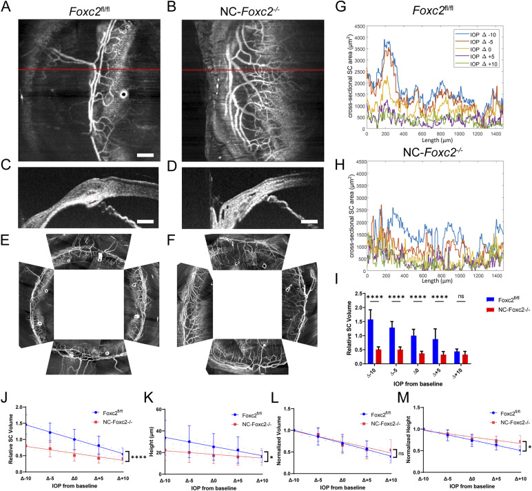
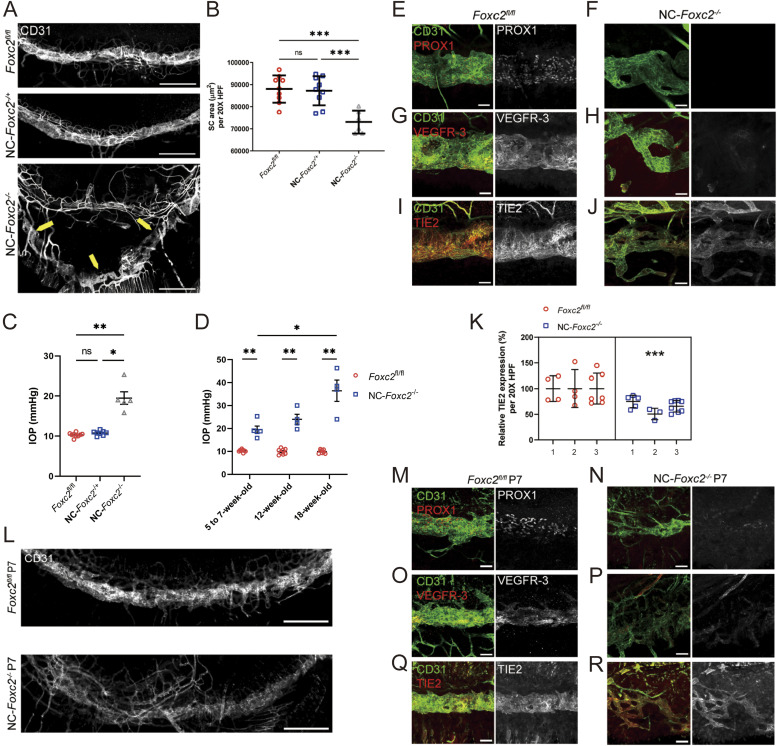
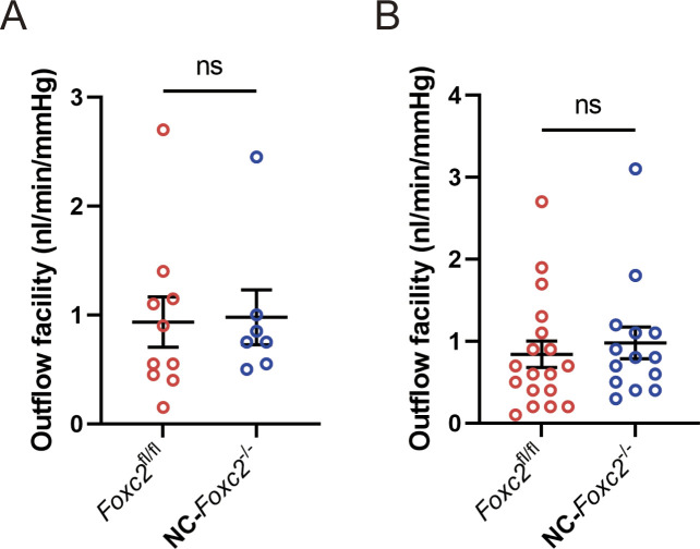
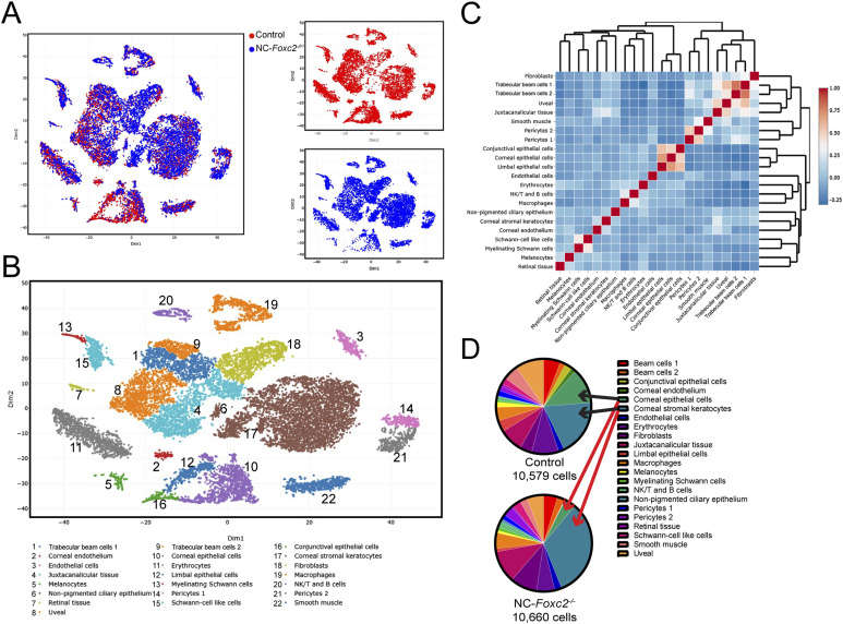
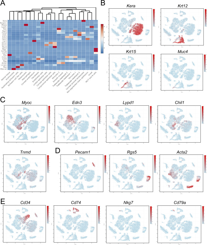
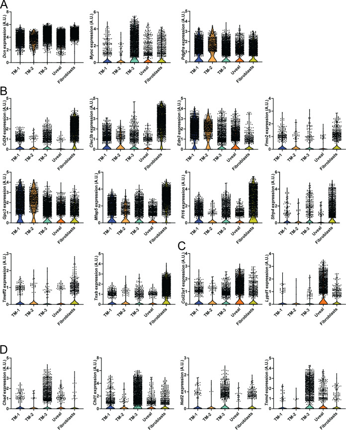
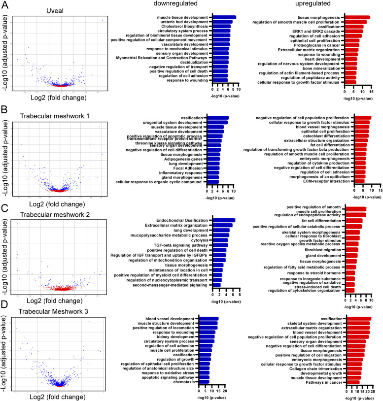
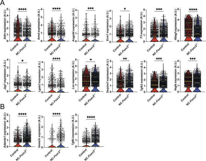
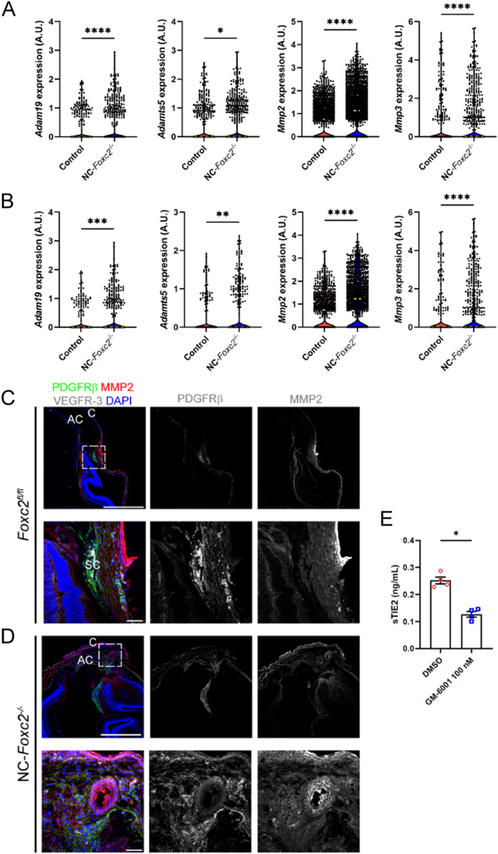
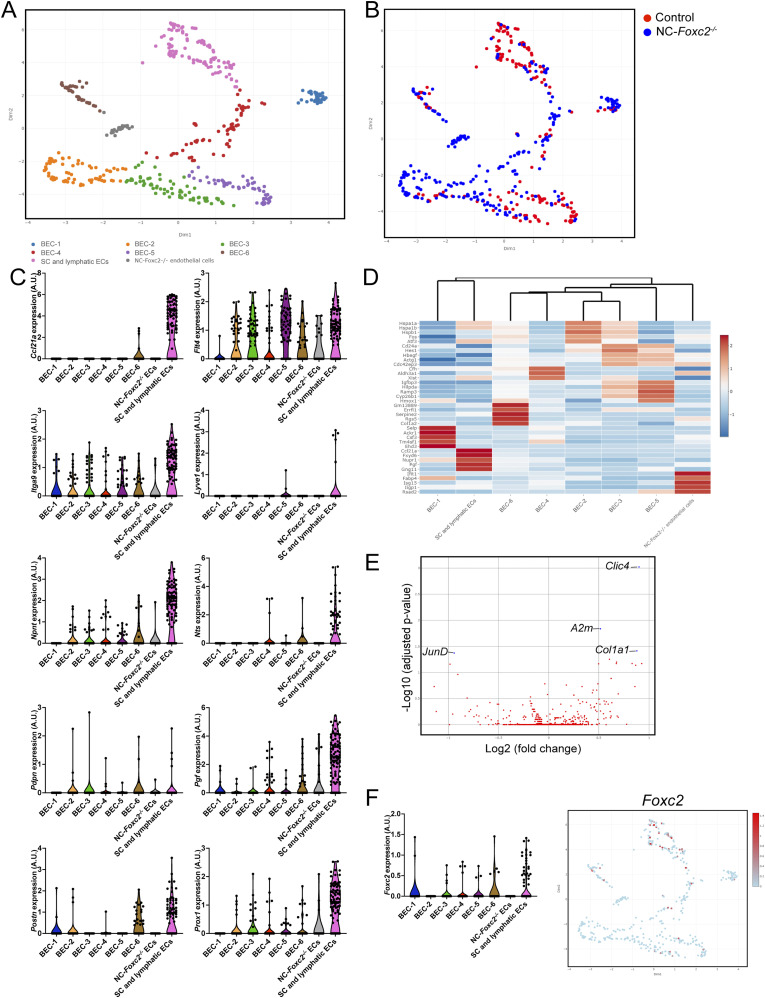
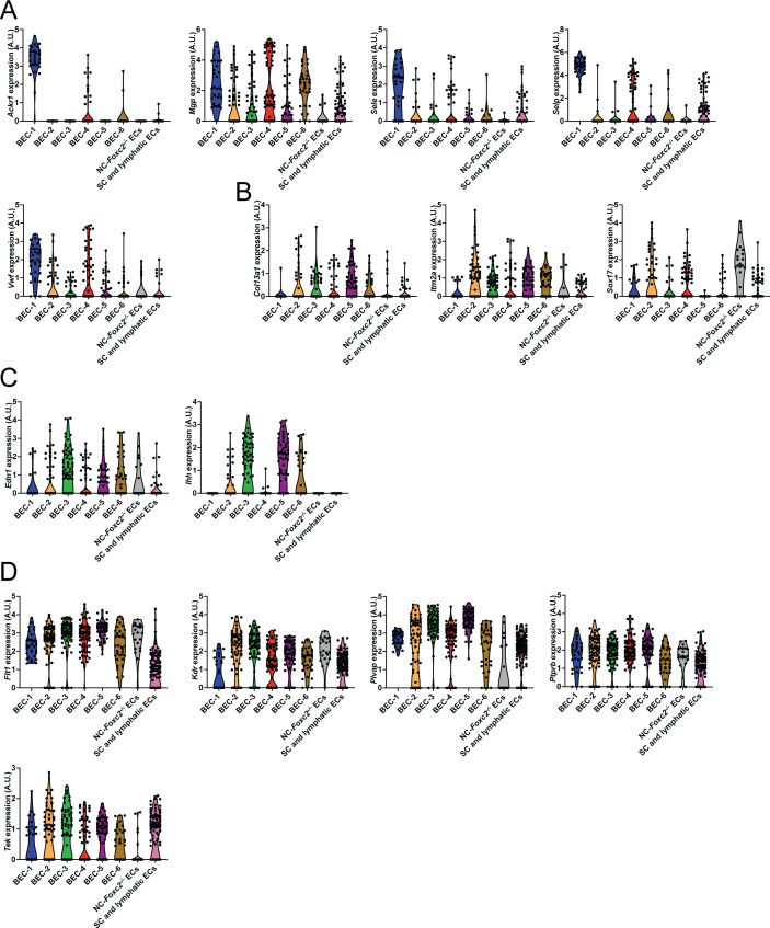
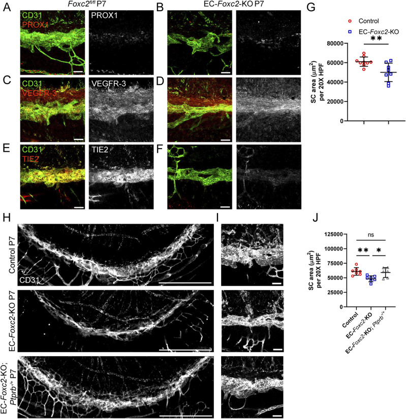
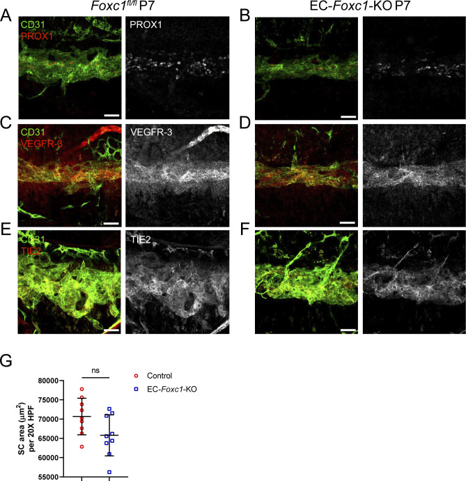
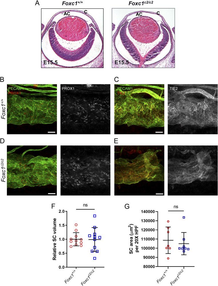
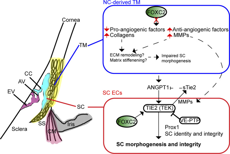
Impaired development and maintenance of Schlemm's canal (SC) are associated with perturbed aqueous humor outflow and intraocular pressure. The angiopoietin (ANGPT)/TIE2 signaling pathway regulates SC development and maintenance, whereas the molecular mechanisms of crosstalk between SC and the neural crest (NC)-derived neighboring tissue, the trabecular meshwork (TM), are poorly understood. Here, we show NC-specific forkhead box ( deletion in mice results in impaired SC morphogenesis, loss of SC identity, and elevated intraocular pressure. Visible-light optical coherence tomography analysis further demonstrated functional impairment of the SC in response to changes in intraocular pressure in NC- mice, suggesting altered TM biomechanics. Single-cell RNA-sequencing analysis identified that this phenotype is predominately characterized by transcriptional changes associated with extracellular matrix organization and stiffness in TM cell clusters, including increased matrix metalloproteinase expression, which can cleave the TIE2 ectodomain to produce soluble TIE2. Moreover, endothelial-specific deletion impaired SC morphogenesis because of reduced TIE2 expression, which was rescued by deleting the TIE2 phosphatase VE-PTP. Thus, Foxc2 is critical in maintaining SC identity and morphogenesis via TM-SC crosstalk.

摘要

Schlemm 管 (SC) 的发育和维持受损与房水流出和眼内压改变有关。血管生成素 (ANGPT)/TIE2 信号通路调节 SC 的发育和维持,而 SC 与神经嵴 (NC) 衍生的相邻组织——小梁网 (TM) 之间的串扰的分子机制尚不清楚。在这里,我们显示 NC 特异性叉头框 (Foxc2) 缺失会导致 SC 形态发生受损、SC 特征丧失和眼内压升高。可见光相干断层扫描分析进一步证明了在 NC- 小鼠中,SC 对眼内压变化的功能受损,提示 TM 生物力学发生改变。单细胞 RNA 测序分析表明,这种表型主要表现为与 TM 细胞簇中细胞外基质组织和硬度相关的转录变化,包括基质金属蛋白酶表达增加,其可裂解 TIE2 细胞外结构域产生可溶性 TIE2。此外,内皮特异性 Foxc2 缺失会因 TIE2 表达减少而损害 SC 形态发生,而 VE-PTP 缺失可挽救这一表型。因此,Foxc2 通过 TM-SC 串扰在维持 SC 特征和形态发生方面至关重要。

https://cdn.ncbi.nlm.nih.gov/pmc/blobs/60cb/10326420/4b8a9e52bb3e/LSA-2022-01721_FigS9.jpg
https://cdn.ncbi.nlm.nih.gov/pmc/blobs/60cb/10326420/528851a7fadd/LSA-2022-01721_FigS1.jpg
https://cdn.ncbi.nlm.nih.gov/pmc/blobs/60cb/10326420/a577e1f4dbb4/LSA-2022-01721_FigS2.jpg
https://cdn.ncbi.nlm.nih.gov/pmc/blobs/60cb/10326420/4083e1d94ea5/LSA-2022-01721_Fig1.jpg
https://cdn.ncbi.nlm.nih.gov/pmc/blobs/60cb/10326420/f6848f617731/LSA-2022-01721_Fig2.jpg
https://cdn.ncbi.nlm.nih.gov/pmc/blobs/60cb/10326420/a404363419c9/LSA-2022-01721_FigS3.jpg
https://cdn.ncbi.nlm.nih.gov/pmc/blobs/60cb/10326420/194a142edbee/LSA-2022-01721_Fig3.jpg
https://cdn.ncbi.nlm.nih.gov/pmc/blobs/60cb/10326420/eb9e997cd2a8/LSA-2022-01721_FigS4.jpg
https://cdn.ncbi.nlm.nih.gov/pmc/blobs/60cb/10326420/bfbeb8c26e68/LSA-2022-01721_FigS5.jpg
https://cdn.ncbi.nlm.nih.gov/pmc/blobs/60cb/10326420/5d241b872576/LSA-2022-01721_Fig4.jpg
https://cdn.ncbi.nlm.nih.gov/pmc/blobs/60cb/10326420/c5050fe589c9/LSA-2022-01721_FigS6.jpg
https://cdn.ncbi.nlm.nih.gov/pmc/blobs/60cb/10326420/1a794f79c74d/LSA-2022-01721_Fig5.jpg
https://cdn.ncbi.nlm.nih.gov/pmc/blobs/60cb/10326420/6796d339e3a9/LSA-2022-01721_Fig6.jpg
https://cdn.ncbi.nlm.nih.gov/pmc/blobs/60cb/10326420/7322bb4074a8/LSA-2022-01721_FigS7.jpg
https://cdn.ncbi.nlm.nih.gov/pmc/blobs/60cb/10326420/5c3dc76e8c25/LSA-2022-01721_Fig7.jpg
https://cdn.ncbi.nlm.nih.gov/pmc/blobs/60cb/10326420/eab6a5fed9aa/LSA-2022-01721_FigS8.jpg
https://cdn.ncbi.nlm.nih.gov/pmc/blobs/60cb/10326420/5ef80f2edbb2/LSA-2022-01721_Fig8.jpg
https://cdn.ncbi.nlm.nih.gov/pmc/blobs/60cb/10326420/185fb69db6ab/LSA-2022-01721_Fig9.jpg
https://cdn.ncbi.nlm.nih.gov/pmc/blobs/60cb/10326420/4b8a9e52bb3e/LSA-2022-01721_FigS9.jpg

相似文献

1
Differential roles of FOXC2 in the trabecular meshwork and Schlemm's canal in glaucomatous pathology.叉头框蛋白 C2 在青光眼病理中的小梁网和施莱姆氏管中的差异作用。
Life Sci Alliance. 2023 Jul 6;6(9). doi: 10.26508/lsa.202201721. Print 2023 Sep.
2
A Histomorphometric and Computational Investigation of the Stabilizing Role of Pectinate Ligaments in the Aqueous Outflow Pathway.一项关于翼状襞在房水流出道中稳定作用的组织形态计量学和计算研究。
J Biomech Eng. 2024 Aug 1;146(8). doi: 10.1115/1.4065164.
3
Schlemm's canal-selective Tie2/TEK knockdown induces sustained ocular hypertension in adult mice.Schlemm 氏管选择性 Tie2/TEK 敲低可诱导成年小鼠持续性眼压升高。
Exp Eye Res. 2024 Nov;248:110114. doi: 10.1016/j.exer.2024.110114. Epub 2024 Oct 3.
4
[Modeling and finite element analysis of human trabecular meshwork outflow pathways].[人小梁网流出途径的建模与有限元分析]
Sheng Wu Yi Xue Gong Cheng Xue Za Zhi. 2025 Jun 25;42(3):585-591. doi: 10.7507/1001-5515.202407037.
5
Modeling the Endothelial Glycocalyx Layer in the Human Conventional Aqueous Outflow Pathway.在人传统房水流出通路中模拟内皮糖萼层。
Cells. 2022 Dec 4;11(23):3925. doi: 10.3390/cells11233925.
6
Prion protein modulates endothelial to mesenchyme-like transition in trabecular meshwork cells: Implications for primary open angle glaucoma.朊病毒蛋白调节小梁细胞的内皮向间质样转化:原发性开角型青光眼的相关影响。
Sci Rep. 2019 Sep 11;9(1):13090. doi: 10.1038/s41598-019-49482-6.
7
Nanotubules and Cellular Communication in Trabecular Meshwork Cells.小梁细胞中的纳米管和细胞通讯。
Methods Mol Biol. 2025;2858:49-62. doi: 10.1007/978-1-0716-4140-8_5.
8
Fibrotic Changes to Schlemm's Canal Endothelial Cells in Glaucoma.青光眼中海绵窦内皮细胞的纤维化改变。
Int J Mol Sci. 2021 Aug 31;22(17):9446. doi: 10.3390/ijms22179446.
9
Digital spatial profiling of segmental outflow regions in trabecular meshwork reveals a role for ADAM15.小梁网节段性流出区域的数字空间分析揭示了 ADAM15 的作用。
PLoS One. 2024 Feb 23;19(2):e0298802. doi: 10.1371/journal.pone.0298802. eCollection 2024.
10
The Effect of Intraocular Pressure Load Boundary on the Biomechanics of the Human Conventional Aqueous Outflow Pathway.眼内压负荷边界对人传统房水流出途径生物力学的影响。
Bioengineering (Basel). 2022 Nov 10;9(11):672. doi: 10.3390/bioengineering9110672.

引用本文的文献

1
Single-Cell Characterization of Anterior Segment Development: Cell Types, Pathways, and Signals Driving Formation of the Trabecular Meshwork and Schlemm's Canal.眼前节发育的单细胞特征:驱动小梁网和施莱姆管形成的细胞类型、途径和信号
bioRxiv. 2025 Aug 21:2025.08.15.670438. doi: 10.1101/2025.08.15.670438.
2
The Advance of Single-Cell RNA Sequencing Applications in Ocular Physiology and Disease Research.单细胞RNA测序在眼生理学和疾病研究中的应用进展
Biomolecules. 2025 Aug 4;15(8):1120. doi: 10.3390/biom15081120.
3
Transcriptomic profiling of Schlemm's canal cells reveals a lymphatic-biased identity and three major cell states.

本文引用的文献

1
Decreased outflow facility and Schlemm's canal defects in a mouse model of glaucoma.青光眼小鼠模型中流出道功能降低和施莱姆氏管缺陷。
Exp Eye Res. 2022 Dec;225:109249. doi: 10.1016/j.exer.2022.109249. Epub 2022 Sep 21.
2
Cellular crosstalk regulates the aqueous humor outflow pathway and provides new targets for glaucoma therapies.细胞串扰调节房水流出通路,并为青光眼治疗提供新靶点。
Nat Commun. 2021 Oct 18;12(1):6072. doi: 10.1038/s41467-021-26346-0.
3
FOXC2 controls adult lymphatic endothelial specialization, function, and gut lymphatic barrier preventing multiorgan failure.
对施莱姆氏管细胞的转录组谱分析揭示了其具有偏向于淋巴的特征,并存在三种主要的细胞状态。
Elife. 2024 Oct 18;13:RP96459. doi: 10.7554/eLife.96459.
4
Schlemm's canal-selective Tie2/TEK knockdown induces sustained ocular hypertension in adult mice.Schlemm 氏管选择性 Tie2/TEK 敲低可诱导成年小鼠持续性眼压升高。
Exp Eye Res. 2024 Nov;248:110114. doi: 10.1016/j.exer.2024.110114. Epub 2024 Oct 3.
5
FOXC1 regulates endothelial CD98 (LAT1/4F2hc) expression in retinal angiogenesis and blood-retina barrier formation.FOXC1 在视网膜血管生成和血视网膜屏障形成中调节内皮细胞 CD98(LAT1/4F2hc)的表达。
Nat Commun. 2024 May 16;15(1):4097. doi: 10.1038/s41467-024-48134-2.
6
Transcriptomic profiling of Schlemm's canal cells reveals a lymphatic-biased identity and three major cell states.施莱姆管细胞的转录组分析揭示了一种偏向淋巴管的特征和三种主要细胞状态。
bioRxiv. 2024 Aug 6:2023.08.31.555823. doi: 10.1101/2023.08.31.555823.
FOXC2 控制成人淋巴管内皮细胞的特化、功能和肠道淋巴管屏障,防止多器官衰竭。
Sci Adv. 2021 Jul 16;7(29). doi: 10.1126/sciadv.abf4335. Print 2021 Jul.
4
Effects of Trabecular Meshwork Width and Schlemm's Canal Area on Intraocular Pressure Reduction in Glaucoma Patients.小梁网宽度和施莱姆氏管面积对青光眼患者眼压降低的影响。
Korean J Ophthalmol. 2021 Aug;35(4):311-317. doi: 10.3341/kjo.2021.0007. Epub 2021 Jun 21.
5
Modeling the effects of glaucoma surgery on intraocular pressure.模拟青光眼手术对眼内压的影响。
Exp Eye Res. 2021 Aug;209:108620. doi: 10.1016/j.exer.2021.108620. Epub 2021 May 25.
6
A single cell atlas of human cornea that defines its development, limbal progenitor cells and their interactions with the immune cells.人类眼角膜单细胞图谱,定义其发育、角膜缘祖细胞及其与免疫细胞的相互作用。
Ocul Surf. 2021 Jul;21:279-298. doi: 10.1016/j.jtos.2021.03.010. Epub 2021 Apr 16.
7
Matrix stiffness primes lymphatic tube formation directed by vascular endothelial growth factor-C.基质硬度促进血管内皮生长因子-C 诱导的淋巴管生成。
FASEB J. 2021 May;35(5):e21498. doi: 10.1096/fj.202002426RR.
8
Epidemiology of Glaucoma: The Past, Present, and Predictions for the Future.青光眼的流行病学:过去、现在及未来预测
Cureus. 2020 Nov 24;12(11):e11686. doi: 10.7759/cureus.11686.
9
A Small Molecule Inhibitor of VE-PTP Activates Tie2 in Schlemm's Canal Increasing Outflow Facility and Reducing Intraocular Pressure.一种小分子 VE-PTP 抑制剂激活小梁网中的 Tie2,增加房水流出效率,降低眼压。
Invest Ophthalmol Vis Sci. 2020 Dec 1;61(14):12. doi: 10.1167/iovs.61.14.12.
10
Trends in prevalence of blindness and distance and near vision impairment over 30 years: an analysis for the Global Burden of Disease Study.30 多年来盲症和远距离及近距离视力损伤流行率的变化趋势:全球疾病负担研究的分析。
Lancet Glob Health. 2021 Feb;9(2):e130-e143. doi: 10.1016/S2214-109X(20)30425-3. Epub 2020 Dec 1.