Hong Kyung Soo, Ahn Heeyun, Lee Kwan Ho, Chung Jin Hong, Shin Kyeong-Cheol, Jin Hyun Jung, Jang Jong Geol, Lee Seok Soo, Jang Min Hye, Ahn June Hong
Division of Pulmonology and Allergy, Department of Internal Medicine, Respiratory Center, Yeungnam University Medical Center, Yeungnam University College of Medicine, Daegu, Republic of Korea.
Department of Thoracic and Cardiovascular Surgery, Yeungnam University Medical Center, Yeungnam University College of Medicine, Daegu, Republic of Korea.
Tuberc Respir Dis (Seoul). 2021 Oct;84(4):282-290. doi: 10.4046/trd.2021.0002. Epub 2021 Jun 24.
Radial probe endobronchial ultrasound-guided transbronchial lung biopsy (RP-EBUS-TBLB) has improved the diagnostic yield of bronchoscopic biopsy of peripheral pulmonary lesions (PPLs). The diagnostic yield and complications of RP-EBUS-TBLB for PPLs vary depending on the technique, such as using a guide sheath (GS) or fluoroscopy. In this study, we investigated the utility of RP-EBUS-TBLB using a GS without fluoroscopy for diagnosing PPLs.
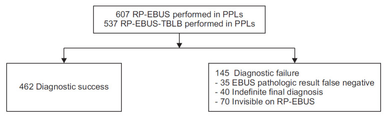
We retrospectively reviewed data from 607 patients who underwent RP-EBUS of PPLs from January 2019 to July 2020. TBLB was performed using RP-EBUS with a GS without fluoroscopy. The diagnostic yield and complications were assessed. Multivariable logistic regression analyses were used to identify factors affecting the diagnostic yields.
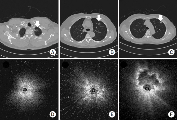
The overall diagnostic accuracy was 76.1% (462/607). In multivariable analyses, the size of the lesion (≥20 mm; odds ratio [OR], 2.06; 95% confidence interval [CI], 1.27-3.33; p=0.003), positive bronchus sign in chest computed tomography (OR, 2.30; 95% CI, 1.40-3.78; p=0.001), a solid lesion (OR, 2.40; 95% CI, 1.31-4.41; p=0.005), and an EBUS image with the probe within the lesion (OR, 6.98; 95% CI, 4.38-11.12; p<0.001) were associated with diagnostic success. Pneumothorax occurred in 2.0% (12/607) of cases and chest tube insertion was required in 0.5% (3/607) of patients.
RP-EBUS-TBLB using a GS without fluoroscopy is a highly accurate diagnostic method in diagnosing PPLs that does not involve radiation exposure and has acceptable complication rates.
径向探头支气管内超声引导下经支气管肺活检(RP-EBUS-TBLB)提高了周围型肺部病变(PPL)支气管镜活检的诊断率。RP-EBUS-TBLB对PPL的诊断率和并发症因技术不同而有所差异,如使用引导鞘(GS)或荧光透视。在本研究中,我们调查了不使用荧光透视的GS辅助RP-EBUS-TBLB对PPL的诊断效用。
我们回顾性分析了2019年1月至2020年7月期间607例行PPL的RP-EBUS患者的数据。使用不配备荧光透视的GS辅助RP-EBUS进行TBLB。评估诊断率和并发症。采用多变量逻辑回归分析确定影响诊断率的因素。
总体诊断准确率为76.1%(462/607)。多变量分析显示,病变大小(≥20 mm;比值比[OR],2.06;95%置信区间[CI],1.27 - 3.33;p = 0.003)、胸部计算机断层扫描中的支气管征阳性(OR,2.30;95% CI,1.40 - 3.78;p = 0.001)、实性病变(OR,2.40;95% CI,1.31 - 4.41;p = 0.005)以及探头位于病变内的EBUS图像(OR,6.98;95% CI,4.38 - 11.12;p < 0.001)与诊断成功相关。气胸发生率为2.0%(12/607),0.5%(3/607)的患者需要插入胸管。
不使用荧光透视的GS辅助RP-EBUS-TBLB是诊断PPL的一种高度准确的诊断方法,不涉及辐射暴露且并发症发生率可接受。