Department of Radiology, Tehran University of Medical Sciences (TUMS), Tehran, Iran.
Department of Radiology, Kabul University of Medical Sciences, Kabul, Afghanistan.
J Med Case Rep. 2021 Jun 24;15(1):324. doi: 10.1186/s13256-021-02945-9.
Fascioliasis is a food-borne hepatobiliary zoonosis caused by Fasciola hepatica and Fasciola gigantica. Human infestations are predominantly seen in developing countries where the disease is endemic, but, due to the increase in international travel rates, hepatic fascioliasis is also appearing in nonendemic areas including Europe and the USA. The clinical and laboratory findings are usually nonspecific. Cross-sectional imaging can be very helpful in the diagnosis of fascioliasis as well as to differentiate it from other liver diseases with a very similar clinical picture. The objectives of this case report are to discuss imaging findings of hepatic fascioliasis and to review the literature.
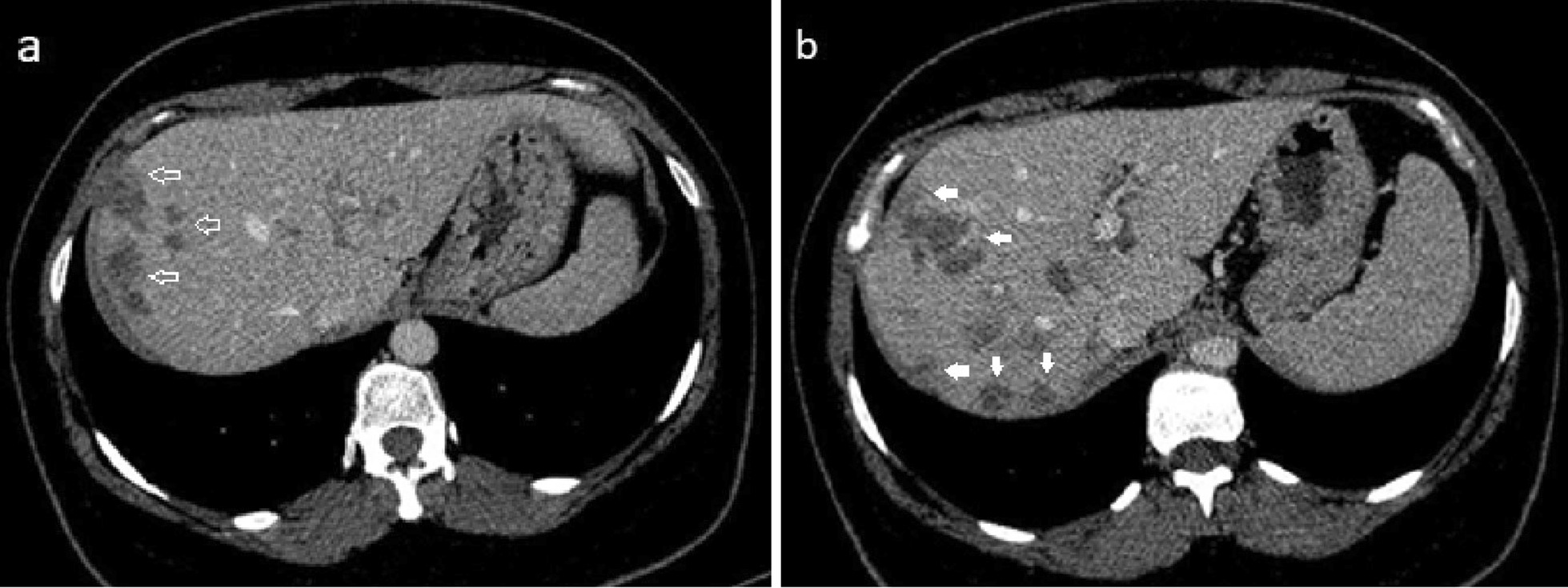
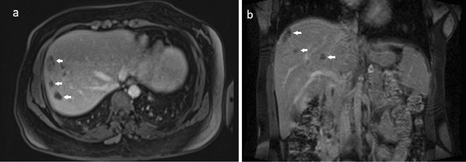
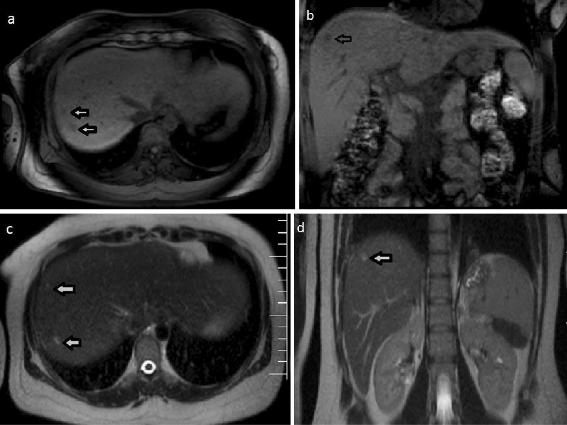
We report the case of a 35-year-old Iranian patient who presented with right upper quadrant pain, low-grade fever, fatigue, and anorexia. The patient had a history of recent travel to the Gilan Province of Iran, almost a month before the onset of symptoms, which is an endemic area of fascioliasis in the country. Laboratory examinations revealed eosinophilia, elevated hepatic enzymes, and slightly raised C-reactive protein. Contrast-enhanced computed tomography of the patient shows clusters of focal ill-defined hypodense lesions with mild peripheral enhancement in the right liver lobe and subcapsular regions. Magnetic resonance imaging of the liver revealed multiple ill-defined lesions of low signal intensity on the T1-weighted image and high signal intensity on the T2-weighted image, extending from the liver capsule into deeper parenchyma toward periportal regions, which shows mild peripheral enhancement on post-contrast images. Imaging-based diagnosis of fascioliasis was made depending on the characteristic distribution of subcapsular tracts/lesions on the above-mentioned imaging, which was then confirmed by serologic tests using enzyme-linked immunosorbent assay. The patient was treated with triclabendazole, showing great clinical improvement, and was eventually discharged in good health condition.
The imaging findings in this case report highlight the importance of cross-sectional imaging for further evaluation of suspected cases of fluke-induced liver disease. The hypothesis of hepatic fascioliasis should be always considered when consistent radiological findings are observed. Clusters of tortuous subcapsular lesions with peripheral contrast enhancement extending into deeper liver parenchyma are characteristic imaging findings that strongly suggest hepatic fascioliasis.
片形吸虫病是一种食源性肝胆人畜共患寄生虫病,由肝片形吸虫和巨片形吸虫引起。人体感染主要见于疾病流行的发展中国家,但由于国际旅行率的增加,包括欧洲和美国在内的非流行地区也出现了肝片形吸虫病。临床和实验室检查结果通常是非特异性的。横断面成像在诊断片形吸虫病以及将其与具有非常相似临床表现的其他肝病区分开来方面非常有帮助。本病例报告的目的是讨论肝片形吸虫病的影像学表现并复习文献。
我们报告了一名 35 岁的伊朗患者的病例,该患者出现右上腹疼痛、低热、乏力和食欲不振。患者最近有前往伊朗吉兰省的旅行史,在症状出现前将近一个月,该国的该地区是片形吸虫病的流行区。实验室检查显示嗜酸性粒细胞增多、肝酶升高和 C 反应蛋白略有升高。患者的增强 CT 显示右肝叶和包膜下区域有簇状局灶性边界不清的低密病灶,伴轻度周边强化。肝脏磁共振成像显示多个边界不清的病灶在 T1 加权像上呈低信号强度,在 T2 加权像上呈高信号强度,从肝包膜向深部实质延伸至门脉周围区域,在增强后图像上显示轻度周边强化。根据上述影像学上特征性的包膜下轨迹/病变分布,做出片形吸虫病的影像学诊断,然后通过酶联免疫吸附试验进行血清学检测来证实。患者接受三氯苯达唑治疗,临床症状显著改善,最终健康状况良好出院。
本病例报告中的影像学表现强调了横断面成像对疑似吸虫病引起的肝病进一步评估的重要性。当观察到一致的放射学发现时,应始终考虑肝片形吸虫病的假说。特征性的影像学表现为簇状迂曲的包膜下病变,伴周边对比增强延伸至深部肝实质,强烈提示肝片形吸虫病。