Mei Youmin, Shen Xiang, Wang Xiaoqian, Zhang Min, Li Qiao, Yan Junyi, Xu Jiali, Xu Yan
Jiangsu Key Laboratory of Oral Diseases, Nanjing Medical University, Nanjing, China.
Department of Periodontology, Nantong Stomatological Hospital, The Affiliated Nantong Stomatological Hospital of Nantong University, Nantong, China.
PeerJ. 2021 Jun 9;9:e11577. doi: 10.7717/peerj.11577. eCollection 2021.
This study aimed to investigate the expression of autophagy-related factors microtubule-associated protein l light chain 3 (LC3) and the apoptosis-related factors BCL2-associated X protein (Bax) and B cell lymphoma-2 (Bcl-2) in the periodontal tissue of experimental diabetic rats. These data were used to explore the potential mechanism in diabetes-induced periodontal tissue lesions.
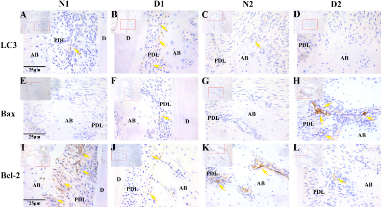
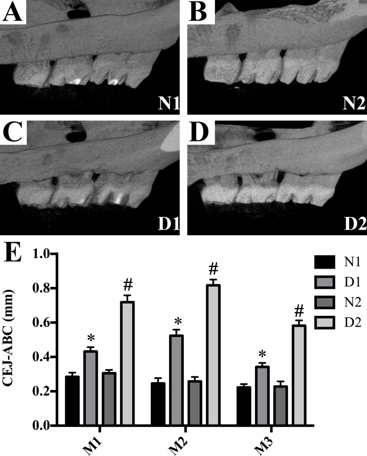
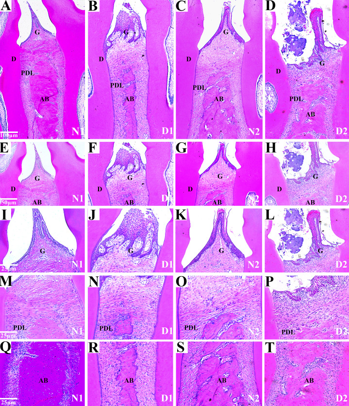
A total of 32 Sprague Dawley (SD) rats were randomly assigned into diabetes (group D, = 16) and control groups (group N, = 16). The diabetic group was induced by intraperitoneal injection of 1% streptozotocin (STZ, 60 mg/kg) and the control group was injected with citrate buffer (0.1mol/L). Rats were sacrificed after 4 and 8 weeks of feeding and collected as D1, N1 groups and D2, N2 groups, and the maxilla were retained for analysis. The changes in periodontal tissue structure were observed by hematoxylin-eosin (HE) staining. The expression and distribution of LC3, Bax and Bcl-2 in the periodontium of the rats was detected by immunohistochemical (SP) staining.
Diabetic rats showed several changes compared to control animals including sparse alveolar bone trabecular structure, loss of the lamina dura and absorption of the local alveolar bone. The positive expression level of LC3 in the gingival epithelial, periodontal ligament and alveolar bone of group D1 was significantly higher than in the N1, N2 and D2 groups ( < 0.05). The level of Bax expression in the group D2 rats was significantly higher than those in the N1, N2 and D1 groups ( < 0.05), while the positive degree of Bcl-2 was significantly lower than those of other groups ( < 0.001). LC3 was negatively correlated with Bax and was irrelevant with Bcl-2; Bcl-2 was not correlated with Bax.
The expression of LC3, Bax and Bcl-2 changes in the periodontal tissue of diabetic rats may indicate that autophagy and apoptotic are involved in the process of periodontal tissue damage in diabetic rats. These changes may be one of the mechanisms of periodontal tissue lesions.
本研究旨在探讨自噬相关因子微管相关蛋白1轻链3(LC3)以及凋亡相关因子BCL2相关X蛋白(Bax)和B细胞淋巴瘤-2(Bcl-2)在实验性糖尿病大鼠牙周组织中的表达情况。利用这些数据来探究糖尿病诱发牙周组织病变的潜在机制。
总共32只Sprague Dawley(SD)大鼠被随机分为糖尿病组(D组,n = 16)和对照组(N组,n = 16)。糖尿病组通过腹腔注射1%链脲佐菌素(STZ,60 mg/kg)诱导产生,对照组则注射柠檬酸盐缓冲液(0.1mol/L)。喂养4周和8周后处死大鼠,分别收集作为D1、N1组以及D2、N2组,并保留上颌骨用于分析。通过苏木精-伊红(HE)染色观察牙周组织结构的变化。采用免疫组织化学(SP)染色检测大鼠牙周组织中LC3、Bax和Bcl-2的表达及分布情况。
与对照动物相比,糖尿病大鼠出现了多种变化,包括牙槽骨小梁结构稀疏、硬骨板缺失以及局部牙槽骨吸收。D1组牙龈上皮、牙周膜和牙槽骨中LC3的阳性表达水平显著高于N1、N2和D2组(P < 0.05)。D2组大鼠中Bax的表达水平显著高于N1、N2和D1组(P < 0.05),而Bcl-2的阳性程度显著低于其他组(P < 0.001)。LC3与Bax呈负相关,与Bcl-2无关;Bcl-2与Bax无相关性。
糖尿病大鼠牙周组织中LC3、Bax和Bcl-2的表达变化可能表明自噬和凋亡参与了糖尿病大鼠牙周组织损伤的过程。这些变化可能是牙周组织病变的机制之一。