Kvernby Sofia, Hult Nafsika Korsavidou, Lindström Elin, Sigfridsson Jonathan, Linder Gustav, Hedberg Jakob, Ahlström Håkan, Bjerner Tomas, Lubberink Mark
Radiology & Nuclear Medicine, Department of Surgical Sciences, Uppsala University, Uppsala, Sweden.
Medical Physics, Uppsala University Hospital, Entrance 85, SE-751 85, Uppsala, Sweden.
Eur J Hybrid Imaging. 2021 Mar 23;5(1):5. doi: 10.1186/s41824-021-00099-x.
Respiratory motion during PET imaging reduces image quality. Data-driven gating (DDG) based on principal component analysis (PCA) can be used to identify respiratory signals. The use of DDG, without need for external devices, would greatly increase the feasibility of using respiratory gating in a routine clinical setting. The objective of this study was to evaluate data-driven gating in relation to external hardware gating and regular static image acquisition on PET-MRI data with respect to SUV and lesion volumes.
Sixteen patients with esophageal or gastroesophageal cancer (Siewert I and II) underwent a 6-min PET scan on a Signa PET-MRI system (GE Healthcare) 1.5-2 h after injection of 4 MBq/kg F-FDG. External hardware gating was done using a respiratory bellow device, and DDG was performed using MotionFree (GE Healthcare). The DDG raw data files and the external hardware-gating raw files were created on a Matlab-based toolbox from the whole 6-min scan LIST-file. For comparison, two 3-min static raw files were created for each patient. Images were reconstructed using TF-OSEM with resolution recovery with 2 iterations, 28 subsets, and 3-mm post filter. SUV and lesion volume were measured in all visible lesions, and noise level was measured in the liver. Paired t-test, linear regression, Pearson correlation, and Bland-Altman analysis were used to investigate difference, correlation, and agreement between the methods.
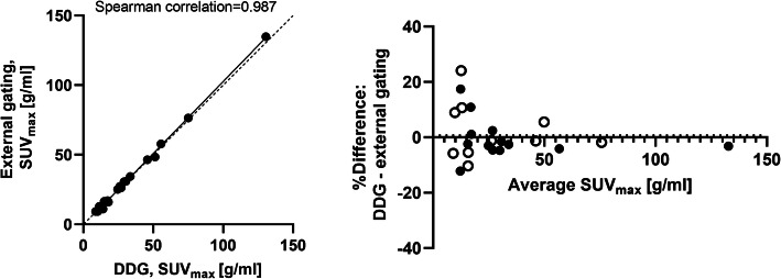
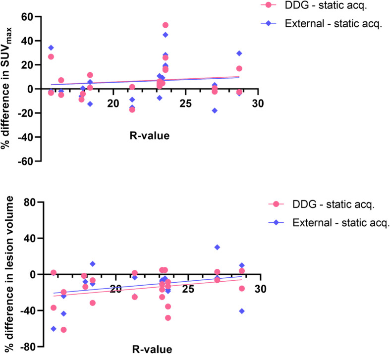
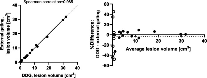
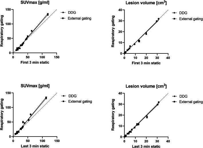
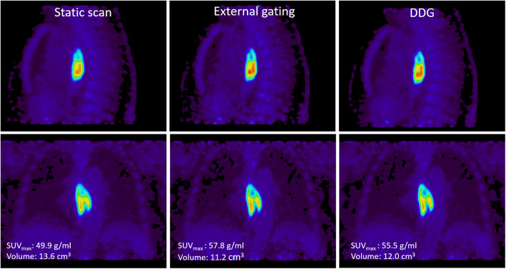
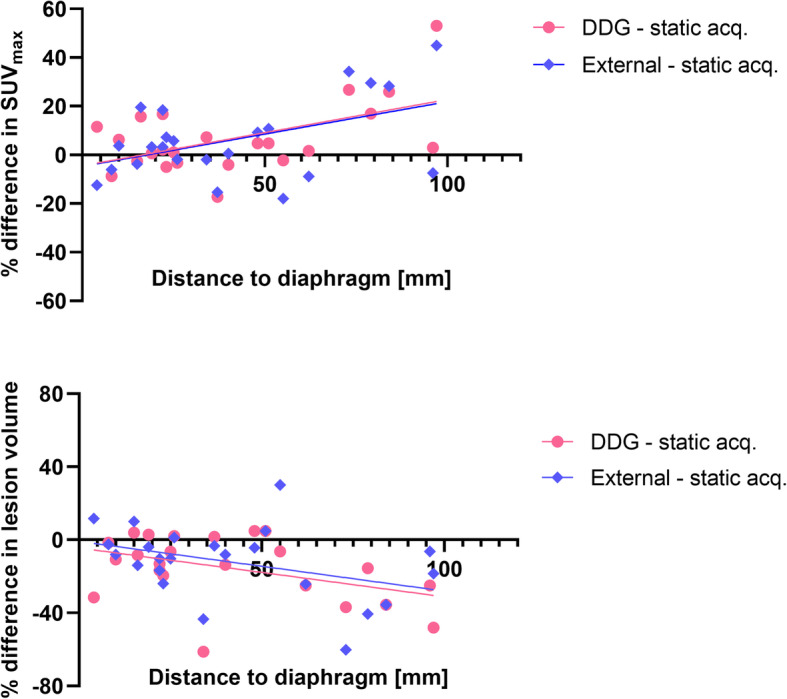
A total number of 30 lesions were included in the study. No significant differences between DDG and external hardware-gating SUV or lesion volumes were found, but the noise level was significantly reduced in the DDG images. Both DDG and external hardware gating demonstrated significantly higher SUV (9.4% for DDG, 10.3% for external hardware gating) and smaller lesion volume (- 5.4% for DDG, - 6.6% for external gating) in comparison with non-gated static images.
Data-driven gating with MotionFree for PET-MRI performed similar to external device gating for esophageal lesions with respect to SUV and lesion volume. Both gating methods significantly increased the SUV and reduced the lesion volume in comparison with non-gated static acquisition. DDG resulted in reduced image noise compared to external device gating and static images.
PET成像过程中的呼吸运动会降低图像质量。基于主成分分析(PCA)的数据驱动门控(DDG)可用于识别呼吸信号。使用DDG无需外部设备,这将大大提高在常规临床环境中使用呼吸门控的可行性。本研究的目的是在SUV和病变体积方面,评估PET-MRI数据中数据驱动门控与外部硬件门控及常规静态图像采集的关系。
16例食管或胃食管癌(Siewert I和II型)患者在注射4 MBq/kg F-FDG后1.5 - 2小时,在Signa PET-MRI系统(GE医疗)上进行6分钟的PET扫描。使用呼吸波纹管装置进行外部硬件门控,使用MotionFree(GE医疗)进行DDG。从整个6分钟扫描的LIST文件中,在基于Matlab的工具箱上创建DDG原始数据文件和外部硬件门控原始文件。为作比较,为每位患者创建两个3分钟的静态原始文件。使用TF-OSEM并进行分辨率恢复,通过2次迭代、28个子集和3毫米后置滤波器重建图像。在所有可见病变中测量SUV和病变体积,在肝脏中测量噪声水平。使用配对t检验、线性回归、Pearson相关性分析和Bland-Altman分析来研究各方法之间的差异、相关性和一致性。
本研究共纳入30个病变。未发现DDG与外部硬件门控的SUV或病变体积之间存在显著差异,但DDG图像中的噪声水平显著降低。与非门控静态图像相比,DDG和外部硬件门控均显示出显著更高的SUV(DDG为9.4%,外部硬件门控为10.3%)和更小的病变体积(DDG为 - 5.4%,外部门控为 - 6.6%)。
对于PET-MRI,使用MotionFree进行数据驱动门控在SUV和病变体积方面与食管病变的外部设备门控效果相似。与非门控静态采集相比,两种门控方法均显著提高了SUV并减小了病变体积。与外部设备门控和静态图像相比,DDG降低了图像噪声。