Attaran Bahareh, Salehi Najmeh, Ghadiri Bahareh, Esmaeili Maryam, Kalateh Shadi, Tashakoripour Mohammad, Eshagh Hosseini Mahmoud, Mohammadi Marjan
HPGC Research Group, Department of Medical Biotechnology, Pasteur Institute of Iran, Tehran, Iran.
Department of Microbiology, Faculty of Biological Sciences, Alzahra University, Tehran, Iran.
Gut Pathog. 2021 Jun 28;13(1):43. doi: 10.1186/s13099-021-00438-0.
Amoxicillin-resistant H. pylori strains are increasing worldwide. To explore the potential resistance mechanisms involved, the 3D structure modeling and access tunnel prediction for penicillin-binding proteins (PBP1A) was performed, based on the Streptococcus pneumoniae, PBP 3D structure. Molecular covalent docking was used to determine the interactions between amoxicillin (AMX) and PBP1A.
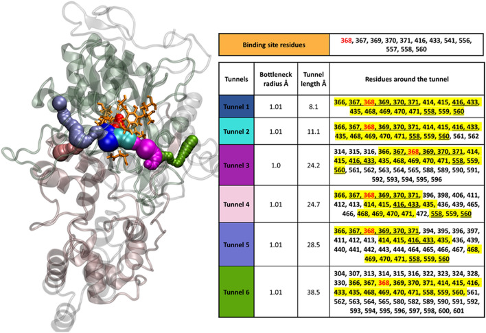
The AMX-Ser368 covalent complex interacts with the binding site residues (Gly367, Ala369, ILE370, Lys371, Tyr416, Ser433, Thr541, Thr556, Gly557, Thr558, and Asn560) of PBP1A, non-covalently. Six tunnel-like structures, accessing the PBP1A binding site, were characterized, using the CAVER algorithm. Tunnel-1 was the ultimate access route, leading to the drug catalytic binding residue (Ser368). This tunnel comprises of eighteen amino acid residues, 8 of which are shared with the drug binding site. Subsequently, to screen the presence of PBP1A mutations, in the binding site and tunnel residues, in our clinical strains, in vitro assays were performed. H. pylori strains, isolated under gastroscopy, underwent AMX susceptibility testing by E-test. Of the 100 clinical strains tested, 4 were AMX-resistant. The transpeptidase domain of the pbp1a gene of these resistant, plus 10 randomly selected AMX-susceptible strains, were amplified and sequenced. Of the amino acids lining the tunnel-1 and binding site residues, three (Ser414Arg, Val469Met and Thr556Ser) substitutions, were detected in 2 of the 4 resistant and none of the sequenced susceptible strains, respectively.
We hypothesize that mutations in amino acid residues lining the binding site and/or tunnel-1, resulting in conformational/spatial changes, may block drug binding to PBP1A and cause AMX resistance.
耐阿莫西林的幽门螺杆菌菌株在全球范围内不断增加。为探究其中潜在的耐药机制,基于肺炎链球菌青霉素结合蛋白(PBP1A)的三维结构进行了三维结构建模及通道预测。采用分子共价对接来确定阿莫西林(AMX)与PBP1A之间的相互作用。
AMX - Ser368共价复合物与PBP1A的结合位点残基(Gly367、Ala369、ILE370、Lys371、Tyr416、Ser433、Thr541、Thr556、Gly557、Thr558和Asn560)发生非共价相互作用。使用CAVER算法对通向PBP1A结合位点的六个隧道样结构进行了表征。通道1是通向药物催化结合残基(Ser368)的最终通道。该通道由18个氨基酸残基组成,其中8个与药物结合位点共有。随后,为筛选临床菌株中PBP1A结合位点和通道残基的突变情况,进行了体外试验。通过胃镜检查分离的幽门螺杆菌菌株采用E-test法进行阿莫西林敏感性测试。在测试的100株临床菌株中,有4株对阿莫西林耐药。对这些耐药菌株以及10株随机选择的阿莫西林敏感菌株的pbp1a基因的转肽酶结构域进行扩增和测序。在通道1和结合位点残基的氨基酸中,分别在4株耐药菌株中的2株以及所有测序的敏感菌株中均未检测到三个(Ser414Arg、Val469Met和Thr556Ser)替换突变。
我们推测结合位点和/或通道1内氨基酸残基的突变导致构象/空间变化,可能会阻止药物与PBP1A结合并导致阿莫西林耐药。