From the Departments of Medical Imaging, Hematology and Clinical Oncology (S.R.T., J.E., M.C.Z.Z.), Gynecology and Obstetrics (C.M.C.), and Pediatrics (A.Y.Y., S.F.B.d.M.N., M.M.M.P.), Ribeirão Preto Medical School, University of São Paulo, Av. Bandeirantes, 3900, Monte Alegre, Ribeirão Preto, São Paulo, Brazil 14049-900; and Department of Radiology, Children's Hospital of Philadelphia, Philadelphia, Pa (S.R.T.).
Radiology. 2021 Sep;300(3):690-698. doi: 10.1148/radiol.2021204150. Epub 2021 Jun 29.
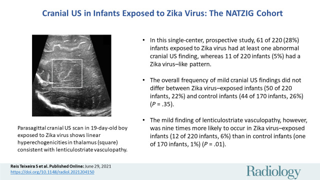
Background Studies addressing neuroimaging findings as primary outcomes of congenital Zika virus infection are variable regarding inclusion criteria and confirmatory laboratory testing. Purpose To investigate cranial US signs of prenatal Zika virus exposure and to describe frequencies of cranial US findings in infants exposed to Zika virus compared to those in control infants. Materials and Methods In this single-center prospective cohort study, participants were enrolled during the December 2015-July 2016 outbreak of Zika virus infection in southeast Brazil (Natural History of Zika Virus Infection in Gestation cohort). Eligibility criteria were available cranial US and laboratory findings of maternal Zika virus infection during pregnancy confirmed with RNA polymerase chain reaction testing (ie, Zika virus-exposed infants). The control group was derived from the Zika in Infants and Pregnancy cohort and consisted of infants born to asymptomatic pregnant women who tested negative for Zika virus infection during pregnancy. Two radiologists who were blinded to the maternal Zika virus infection status independently reviewed cranial US scans from both groups and categorized them as normal findings, Zika virus-like pattern, or mild findings. Associations between cranial US findings and prenatal Zika virus exposure were assessed with univariable analysis. Results Two hundred twenty Zika virus-exposed infants (mean age, 53.3 days ± 71.1 [standard deviation]; 113 boys) and born to 219 mothers infected with Zika virus were included in this study and compared with 170 control infants (mean age, 45.6 days ± 45.8; 102 boys). Eleven of the 220 Zika virus-exposed infants (5%), but no control infants, had a Zika virus-like pattern at cranial US. No difference in frequency of mild findings was observed between the groups (50 of 220 infants [23%] vs 44 of 170 infants [26%], respectively; = .35). The mild finding of lenticulostriate vasculopathy, however, was nine times more frequent in Zika virus-exposed infants (12 of 220 infants, 6%) than in control infants (one of 170 infants, 1%) ( = .01). Conclusion Lenticulostriate vasculopathy was more common after prenatal exposure to Zika virus, even in infants with normal head size, despite otherwise overall similar frequency of mild cranial US findings in Zika virus-exposed infants and in control infants. © RSNA, 2021 . See also the editorial by Benson in this issue.
背景 针对先天性寨卡病毒感染的神经影像学发现作为主要结果的研究,其纳入标准和确证性实验室检测存在差异。 目的 研究产前寨卡病毒暴露的颅超声征象,并描述与对照婴儿相比,暴露于寨卡病毒的婴儿的颅超声发现的频率。 材料与方法 在这项单中心前瞻性队列研究中,参与者于 2015 年 12 月至 2016 年 7 月巴西东南部寨卡病毒感染爆发期间(妊娠期间寨卡病毒感染的自然史队列)入组。纳入标准为有颅超声和实验室检查结果,并经 RNA 聚合酶链反应检测证实母亲妊娠期间感染寨卡病毒(即寨卡病毒暴露婴儿)。对照组来自寨卡病毒与妊娠队列,由妊娠期间寨卡病毒感染检测阴性的无症状孕妇所生婴儿组成。两位对母亲寨卡病毒感染状态不知情的放射科医生独立审查了两组的颅超声扫描,并将其分类为正常发现、寨卡病毒样模式或轻度发现。使用单变量分析评估颅超声发现与产前寨卡病毒暴露之间的关系。 结果 本研究纳入了 220 名寨卡病毒暴露婴儿(平均年龄,53.3 天±71.1[标准差];113 名男婴)和 219 名感染寨卡病毒的母亲,并与 170 名对照婴儿(平均年龄,45.6 天±45.8;102 名男婴)进行了比较。在 220 名寨卡病毒暴露婴儿中,有 11 名(5%)出现了寨卡病毒样模式,而对照组中无一例出现这种模式。两组轻度发现的频率无差异(分别为 220 名婴儿中有 50 名[23%]和 170 名婴儿中有 44 名[26%]; =.35)。然而,在寨卡病毒暴露婴儿中,纹状体动脉病的轻度表现更为常见(220 名婴儿中有 12 名,6%),而在对照婴儿中仅 1 名(1%)( =.01)。 结论 即使在头围大小正常的婴儿中,产前暴露于寨卡病毒后纹状体动脉病也更为常见,尽管暴露于寨卡病毒的婴儿和对照婴儿的颅超声轻度发现总体频率相似。 版权所有©2021 RSNA。 请同时参见本期 Benson 编辑的社论。