• 文献检索
  • 文档翻译
  • 深度研究
  • 学术资讯
  • Suppr Zotero 插件Zotero 插件
  • 邀请有礼
  • 套餐&价格
  • 历史记录
应用&插件
Suppr Zotero 插件Zotero 插件浏览器插件Mac 客户端Windows 客户端微信小程序
定价
高级版会员购买积分包购买API积分包
服务
文献检索文档翻译深度研究API 文档MCP 服务
关于我们
关于 Suppr公司介绍联系我们用户协议隐私条款
关注我们

Suppr 超能文献

核心技术专利:CN118964589B侵权必究
粤ICP备2023148730 号-1Suppr @ 2026

文献检索

告别复杂PubMed语法,用中文像聊天一样搜索,搜遍4000万医学文献。AI智能推荐,让科研检索更轻松。

立即免费搜索

文件翻译

保留排版,准确专业,支持PDF/Word/PPT等文件格式,支持 12+语言互译。

免费翻译文档

深度研究

AI帮你快速写综述,25分钟生成高质量综述,智能提取关键信息,辅助科研写作。

立即免费体验

基于荧光蛋白的自噬生物传感器

Fluorescent Protein-Based Autophagy Biosensors.

作者信息

Kim Heejung, Seong Jihye

机构信息

Brain Science Institute, Korea Institute of Science and Technology (KIST), Seoul 02792, Korea.

Department of Converging Science and Technology, Kyung Hee University, Seoul 02453, Korea.

出版信息

Materials (Basel). 2021 Jun 2;14(11):3019. doi: 10.3390/ma14113019.

DOI:10.3390/ma14113019
PMID:34199451
原文链接:https://pmc.ncbi.nlm.nih.gov/articles/PMC8199620/
Abstract

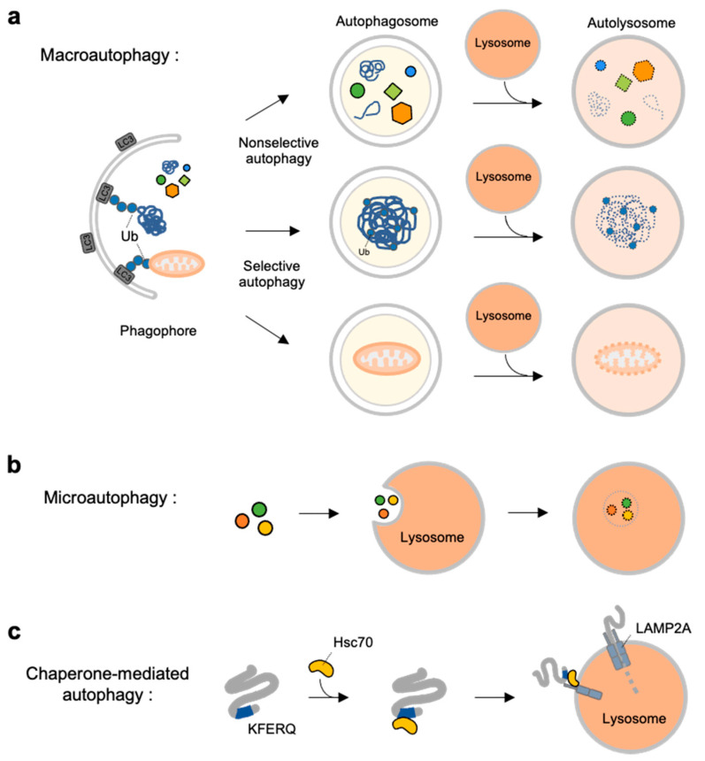
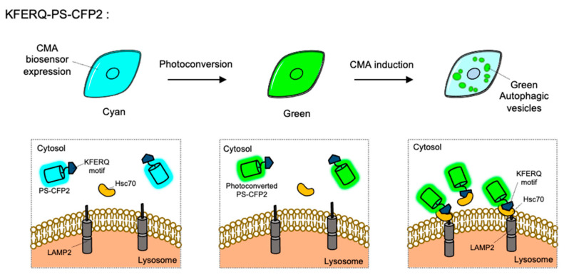
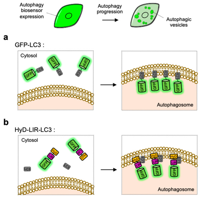
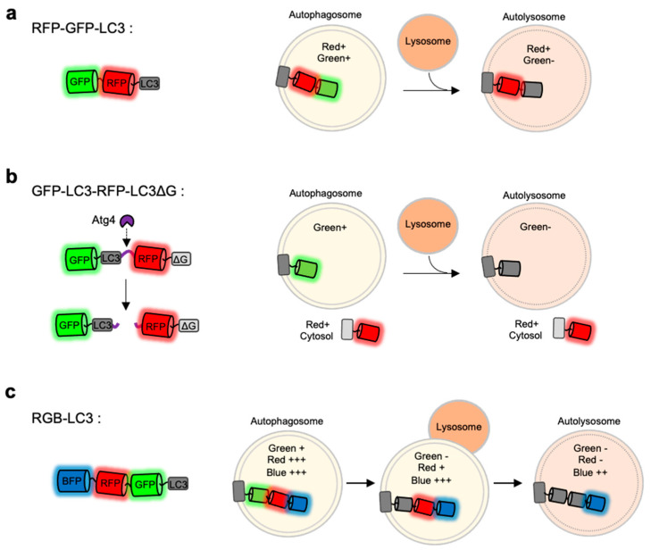
Autophagy is an essential cellular process of self-degradation for dysfunctional or unnecessary cytosolic constituents and organelles. Dysregulation of autophagy is thus involved in various diseases such as neurodegenerative diseases. To investigate the complex process of autophagy, various biochemical, chemical assays, and imaging methods have been developed. Here we introduce various methods to study autophagy, in particular focusing on the review of designs, principles, and limitations of the fluorescent protein (FP)-based autophagy biosensors. Different physicochemical properties of FPs, such as pH-sensitivity, stability, brightness, spectral profile, and fluorescence resonance energy transfer (FRET), are considered to design autophagy biosensors. These FP-based biosensors allow for sensitive detection and real-time monitoring of autophagy progression in live cells with high spatiotemporal resolution. We also discuss future directions utilizing an optobiochemical strategy to investigate the in-depth mechanisms of autophagy. These cutting-edge technologies will further help us to develop the treatment strategies of autophagy-related diseases.

摘要

自噬是一种重要的细胞自我降解过程,用于处理功能失调或不必要的胞质成分和细胞器。因此,自噬失调与多种疾病有关,如神经退行性疾病。为了研究自噬的复杂过程,人们开发了各种生化、化学检测方法和成像方法。在这里,我们介绍各种研究自噬的方法,特别侧重于对基于荧光蛋白(FP)的自噬生物传感器的设计、原理和局限性的综述。在设计自噬生物传感器时,会考虑荧光蛋白的不同物理化学性质,如pH敏感性、稳定性、亮度、光谱特征和荧光共振能量转移(FRET)。这些基于荧光蛋白的生物传感器能够以高时空分辨率在活细胞中灵敏地检测和实时监测自噬进程。我们还讨论了利用光生物化学策略深入研究自噬机制的未来方向。这些前沿技术将进一步帮助我们制定自噬相关疾病的治疗策略。

https://cdn.ncbi.nlm.nih.gov/pmc/blobs/c2f7/8199620/89125e262a6f/materials-14-03019-g005.jpg
https://cdn.ncbi.nlm.nih.gov/pmc/blobs/c2f7/8199620/3e26d4ea10cd/materials-14-03019-g001.jpg
https://cdn.ncbi.nlm.nih.gov/pmc/blobs/c2f7/8199620/bf3e7f379796/materials-14-03019-g002.jpg
https://cdn.ncbi.nlm.nih.gov/pmc/blobs/c2f7/8199620/016110d8c085/materials-14-03019-g003.jpg
https://cdn.ncbi.nlm.nih.gov/pmc/blobs/c2f7/8199620/2dfc4229175c/materials-14-03019-g004.jpg
https://cdn.ncbi.nlm.nih.gov/pmc/blobs/c2f7/8199620/89125e262a6f/materials-14-03019-g005.jpg

相似文献

1
Fluorescent Protein-Based Autophagy Biosensors.基于荧光蛋白的自噬生物传感器
Materials (Basel). 2021 Jun 2;14(11):3019. doi: 10.3390/ma14113019.
2
Genetically Encoded Biosensors Based on Fluorescent Proteins.基于荧光蛋白的基因编码生物传感器。
Sensors (Basel). 2021 Jan 25;21(3):795. doi: 10.3390/s21030795.
3
A Guide to Fluorescent Protein FRET Pairs.荧光蛋白荧光共振能量转移对指南。
Sensors (Basel). 2016 Sep 14;16(9):1488. doi: 10.3390/s16091488.
4
Biosensors; a novel concept in real-time detection of autophagy.生物传感器;实时检测自噬的新概念。
Biosens Bioelectron. 2024 Jun 15;254:116204. doi: 10.1016/j.bios.2024.116204. Epub 2024 Mar 16.
5
Fluorescence proteins, live-cell imaging, and mechanobiology: seeing is believing.荧光蛋白、活细胞成像与力学生物学:眼见为实。
Annu Rev Biomed Eng. 2008;10:1-38. doi: 10.1146/annurev.bioeng.010308.161731.
6
Analysis of autophagosome formation using lentiviral biosensors for live fluorescent cellular imaging.使用慢病毒生物传感器进行活细胞荧光成像分析自噬体形成
Methods Mol Biol. 2015;1219:157-69. doi: 10.1007/978-1-4939-1661-0_12.
7
Unravelling molecular dynamics in living cells: Fluorescent protein biosensors for cell biology.解析活细胞中的分子动力学:用于细胞生物学的荧光蛋白生物传感器
J Microsc. 2025 May;298(2):123-184. doi: 10.1111/jmi.13270. Epub 2024 Feb 15.
8
Designs and applications of fluorescent protein-based biosensors.基于荧光蛋白的生物传感器的设计与应用。
Curr Opin Chem Biol. 2010 Feb;14(1):30-6. doi: 10.1016/j.cbpa.2009.09.033. Epub 2009 Nov 11.
9
Fluorescent Biosensors Based on Single-Molecule Counting.基于单分子计数的荧光生物传感器。
Acc Chem Res. 2016 Sep 20;49(9):1722-30. doi: 10.1021/acs.accounts.6b00237. Epub 2016 Sep 1.
10
Genetically encoded FRET-based biosensors for multiparameter fluorescence imaging.用于多参数荧光成像的基于荧光共振能量转移的基因编码生物传感器。
Curr Opin Biotechnol. 2009 Feb;20(1):19-27. doi: 10.1016/j.copbio.2009.01.003. Epub 2009 Feb 14.

引用本文的文献

1
Image-based temporal profiling of autophagy-related phenotypes.基于图像的自噬相关表型的时间剖析。
Autophagy Rep. 2025;4(1). doi: 10.1080/27694127.2025.2484835. Epub 2025 Apr 8.
2
Quantitative profiling pH heterogeneity of acidic endolysosomal compartments using fluorescence lifetime imaging microscopy.使用荧光寿命成像显微镜对酸性内溶酶体区室的pH异质性进行定量分析。
Mol Biol Cell. 2025 Mar 1;36(3):br8. doi: 10.1091/mbc.E23-06-0220. Epub 2025 Jan 29.
3
Valosin-Containing Protein (VCP): A Review of Its Diverse Molecular Functions and Clinical Phenotypes.

本文引用的文献

1
Optobiochemistry: Genetically Encoded Control of Protein Activity by Light.光遗传学:通过光遗传控制蛋白质活性
Annu Rev Biochem. 2021 Jun 20;90:475-501. doi: 10.1146/annurev-biochem-072420-112431. Epub 2021 Mar 29.
2
pHmScarlet is a pH-sensitive red fluorescent protein to monitor exocytosis docking and fusion steps.pHmScarlet 是一种对 pH 值敏感的红色荧光蛋白,可用于监测胞吐作用的对接和融合步骤。
Nat Commun. 2021 Mar 3;12(1):1413. doi: 10.1038/s41467-021-21666-7.
3
Genetically Encoded Biosensors Based on Fluorescent Proteins.基于荧光蛋白的基因编码生物传感器。
泛素结合酶 VCP(Valosin-Containing Protein):分子功能与临床表型多样性综述。
Int J Mol Sci. 2024 May 22;25(11):5633. doi: 10.3390/ijms25115633.
4
Biosensors; a novel concept in real-time detection of autophagy.生物传感器;实时检测自噬的新概念。
Biosens Bioelectron. 2024 Jun 15;254:116204. doi: 10.1016/j.bios.2024.116204. Epub 2024 Mar 16.
5
Protein aggregation and neurodegenerative disease: Structural outlook for the novel therapeutics.蛋白质聚集与神经退行性疾病:新型疗法的结构展望
Proteins. 2023 Aug 2. doi: 10.1002/prot.26561.
6
Natural-Product-Mediated Autophagy in the Treatment of Various Liver Diseases.天然产物介导的自噬在治疗各种肝脏疾病中的作用。
Int J Mol Sci. 2022 Dec 1;23(23):15109. doi: 10.3390/ijms232315109.
Sensors (Basel). 2021 Jan 25;21(3):795. doi: 10.3390/s21030795.
4
Mitochondrial Dysfunction and Mitophagy in Parkinson's Disease: From Mechanism to Therapy.线粒体功能障碍与帕金森病中的自噬:从机制到治疗。
Trends Biochem Sci. 2021 Apr;46(4):329-343. doi: 10.1016/j.tibs.2020.11.007. Epub 2020 Dec 13.
5
Visualization of Autophagy Progression by a Red-Green-Blue Autophagy Sensor.利用红-绿-蓝自噬传感器观察自噬进程
ACS Sens. 2020 Dec 24;5(12):3850-3861. doi: 10.1021/acssensors.0c00809. Epub 2020 Dec 2.
6
Degradation and Transmission of Tau by Autophagic-Endolysosomal Networks and Potential Therapeutic Targets for Tauopathy.自噬-内溶酶体网络对Tau蛋白的降解与传递及Tau蛋白病的潜在治疗靶点
Front Mol Neurosci. 2020 Oct 16;13:586731. doi: 10.3389/fnmol.2020.586731. eCollection 2020.
7
The fluorescence toolbox for visualizing autophagy.用于可视化自噬的荧光工具盒。
Chem Soc Rev. 2020 Nov 21;49(22):8354-8389. doi: 10.1039/d0cs00913j. Epub 2020 Nov 6.
8
Autophagy in Human Diseases.人类疾病中的自噬
N Engl J Med. 2020 Oct 15;383(16):1564-1576. doi: 10.1056/NEJMra2022774.
9
PEG-Ceramide Nanomicelles Induce Autophagy and Degrade Tau Proteins in N2a Cells.PEG-神经酰胺纳米胶束诱导 N2a 细胞自噬并降解 Tau 蛋白。
Int J Nanomedicine. 2020 Sep 11;15:6779-6789. doi: 10.2147/IJN.S258311. eCollection 2020.
10
Microautophagy - distinct molecular mechanisms handle cargoes of many sizes.微自噬——不同的分子机制处理不同大小的货物。
J Cell Sci. 2020 Sep 9;133(17):jcs246322. doi: 10.1242/jcs.246322.